6MG6 | pdb_00006mg6

Crystal structure of carbon-nitrogen hydrolase from Helicobacter pylori G27


Experimental Data Snapshot

  • Method: X-RAY DIFFRACTION
  • Resolution: 2.10 Å
  • R-Value Free: 
    0.208 (Depositor), 0.210 (DCC) 
  • R-Value Work: 
    0.168 (Depositor), 0.170 (DCC) 
  • R-Value Observed: 
    0.169 (Depositor) 

Starting Model: experimental
View more details

wwPDB Validation   3D Report Full Report


This is version 1.3 of the entry. See complete history


Literature

Crystal structure of carbon-nitrogen hydrolase from Helicobacter pylori G27.

Srivastava, A.Ayanlade, J.P.Suleiman, L.Udell, H.Abendroth, J.Lorimer, D.J.Edwards, T.E.Staker, B.L.Zigweid, R.Subramanian, S.Myler, P.J.Chakafana, G.Asojo, O.A.

(2026) Acta Crystallogr F Struct Biol Commun 

  • DOI: https://doi.org/10.1107/S2053230X26001330
  • Primary Citation of Related Structures:  
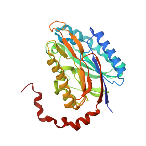
    6MG6

  • PubMed Abstract: 

    Carbon-nitrogen hydrolases (CNHs) are members of the diverse nitrilase superfamily of enzymes that facilitate cellular adaptation to environmental stress by metabolizing nitrogen, detoxifying xenobiotics and catabolizing environmentally derived metabolites. Helicobacter pylori CNH (HpCNH) may contribute to metabolic flexibility under acid stress, detoxification of reactive nitrogen species or nutrient scavenging in the nutrient-limited gastric environment. Here, we report the 2.1 Å resolution crystal structure of a CNH from H. pylori strain G27 (PDB entry 6mg6). HpCNH adopts the characteristic nitrilase-superfamily αββα-sandwich core and contains the conserved catalytic cysteine typical of enzymatically active CNHs. The overall structure and active site of HpCNH are most similar to those of carbamoylputrescine amidohydrolase from the plant Medicago truncatula. Despite structural variations in loop regions, including near the active site, HpCNH retains the key residues required to bind putrescine and the prototypical N-carbamoylputrescine amidase active site.


  • Organizational Affiliation
    • California Institute of Technology, 1200 East California Boulevard, Pasadena, CA 91125, USA.

Macromolecules
Find similar proteins by:  (by identity cutoff)  |  3D Structure
Entity ID: 1
MoleculeChains Sequence LengthOrganismDetailsImage
Carbon-nitrogen hydrolase
A, B, C, D
300Helicobacter pylori G27Mutation(s): 0 
Gene Names: HPG27_713
UniProt
Find proteins for B5Z7B9 (Helicobacter pylori (strain G27))
Explore B5Z7B9 
Go to UniProtKB:  B5Z7B9
Entity Groups  
Sequence Clusters30% Identity50% Identity70% Identity90% Identity95% Identity100% Identity
UniProt GroupB5Z7B9
Sequence Annotations
Expand
  • Reference Sequence
Small Molecules
Ligands 1 Unique
IDChains Name / Formula / InChI Key2D Diagram3D Interactions
SO4
Query on SO4

Download Ideal Coordinates CCD File 
E [auth A]SULFATE ION
O4 S
QAOWNCQODCNURD-UHFFFAOYSA-L
Experimental Data & Validation

Experimental Data

  • Method: X-RAY DIFFRACTION
  • Resolution: 2.10 Å
  • R-Value Free:  0.208 (Depositor), 0.210 (DCC) 
  • R-Value Work:  0.168 (Depositor), 0.170 (DCC) 
  • R-Value Observed: 0.169 (Depositor) 
Space Group: P 21 21 2
Unit Cell:
Length ( Å )Angle ( ˚ )
a = 137.58α = 90
b = 91.38β = 90
c = 95.04γ = 90
Software Package:
Software NamePurpose
XDSdata reduction
XSCALEdata scaling
PHENIXrefinement
PDB_EXTRACTdata extraction
PHASERphasing
PARROTphasing
Cootmodel building
ARP/wARPmodel building

Structure Validation

View Full Validation Report


Entry History 

Deposition Data

Revision History  (Full details and data files)

  • Version 1.0: 2018-09-26
    Type: Initial release
  • Version 1.1: 2024-03-13
    Changes: Data collection, Database references, Refinement description
  • Version 1.2: 2024-04-03
    Changes: Refinement description
  • Version 1.3: 2026-03-04
    Changes: Database references, Structure summary