6MI2

Structure of the human 4-1BB / Utomilumab Fab complex


Experimental Data Snapshot

  • Method: X-RAY DIFFRACTION
  • Resolution: 2.72 Å
  • R-Value Free: 
    0.278 (Depositor), 0.282 (DCC) 
  • R-Value Work: 
    0.238 (Depositor), 0.244 (DCC) 
  • R-Value Observed: 
    0.240 (Depositor) 

wwPDB Validation   3D Report Full Report


Ligand Structure Quality Assessment 

Created with Raphaël 2.3.0Worse 01 BetterLigand structure goodness of fit to experimental dataBest fitted NAGClick on this verticalbar to view details

This is version 2.1 of the entry. See complete history


Literature

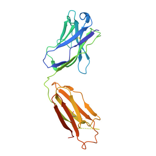
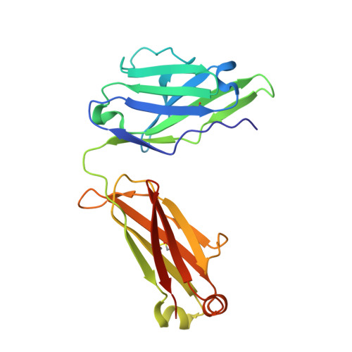
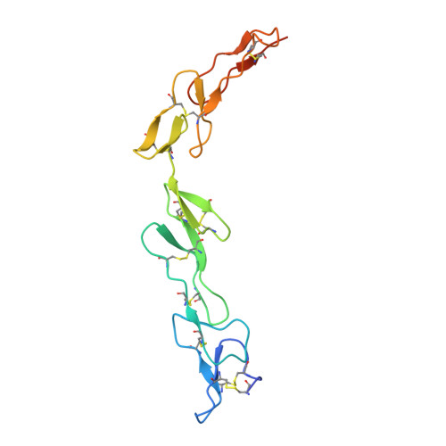
Structure of the 4-1BB/4-1BBL complex and distinct binding and functional properties of utomilumab and urelumab.

Chin, S.M.Kimberlin, C.R.Roe-Zurz, Z.Zhang, P.Xu, A.Liao-Chan, S.Sen, D.Nager, A.R.Oakdale, N.S.Brown, C.Wang, F.Yang, Y.Lindquist, K.Yeung, Y.A.Salek-Ardakani, S.Chaparro-Riggers, J.

(2018) Nat Commun 9: 4679-4679

  • DOI: https://doi.org/10.1038/s41467-018-07136-7
  • Primary Citation of Related Structures:  
    6MGE, 6MHR, 6MI2

  • PubMed Abstract: 

    4-1BB (CD137, TNFRSF9) is an inducible costimulatory receptor expressed on activated T cells. Clinical trials of two agonist antibodies, utomilumab (PF-05082566) and urelumab (BMS-663513), are ongoing in multiple cancer indications, and both antibodies demonstrate distinct activities in the clinic. To understand these differences, we solved structures of the human 4-1BB/4-1BBL complex, the 4-1BBL trimer alone, and 4-1BB bound to utomilumab or urelumab. The 4-1BB/4-1BBL complex displays a unique interaction between receptor and ligand when compared with other TNF family members. Furthermore, our ligand-only structure differs from previously published data. Utomilumab, a ligand-blocking antibody, binds 4-1BB between CRDs 3 and 4. In contrast, urelumab binds 4-1BB CRD-1, away from the ligand binding site. Finally, cell-based assays demonstrate utomilumab is a milder agonist than urelumab. Collectively, our data provide a deeper understanding of the 4-1BB signaling complex, providing a template for future development of next generation 4-1BB targeted biologics.


  • Organizational Affiliation

    Cancer Immunology Discovery, Pfizer Inc., 230 E. Grand Ave, South San Francisco, CA, 94080, USA. shermanmichael.chin@pfizer.com.


Macromolecules
Find similar proteins by:  (by identity cutoff)  |  3D Structure
Entity ID: 1
MoleculeChains Sequence LengthOrganismDetailsImage
Utomilumab Fab heavy chainA,
C [auth D]
230Homo sapiensMutation(s): 0 
Entity Groups  
Sequence Clusters30% Identity50% Identity70% Identity90% Identity95% Identity100% Identity
Sequence Annotations
Expand
  • Reference Sequence
Find similar proteins by:  (by identity cutoff)  |  3D Structure
Entity ID: 2
MoleculeChains Sequence LengthOrganismDetailsImage
Utomilumab Fab lambda chainB,
D [auth E]
214Homo sapiensMutation(s): 0 
Entity Groups  
Sequence Clusters30% Identity50% Identity70% Identity90% Identity95% Identity100% Identity
Sequence Annotations
Expand
  • Reference Sequence
Find similar proteins by:  (by identity cutoff)  |  3D Structure
Entity ID: 3
MoleculeChains Sequence LengthOrganismDetailsImage
Tumor necrosis factor receptor superfamily member 9E [auth F],
F [auth C]
145Homo sapiensMutation(s): 0 
Gene Names: TNFRSF9CD137ILA
UniProt & NIH Common Fund Data Resources
Find proteins for Q07011 (Homo sapiens)
Explore Q07011 
Go to UniProtKB:  Q07011
PHAROS:  Q07011
GTEx:  ENSG00000049249 
Entity Groups  
Sequence Clusters30% Identity50% Identity70% Identity90% Identity95% Identity100% Identity
UniProt GroupQ07011
Glycosylation
Glycosylation Sites: 1Go to GlyGen: Q07011-1
Sequence Annotations
Expand
  • Reference Sequence
Oligosaccharides

Help

Entity ID: 4
MoleculeChains Length2D Diagram Glycosylation3D Interactions
alpha-L-fucopyranose-(1-6)-2-acetamido-2-deoxy-beta-D-glucopyranose
G
2N-Glycosylation
Glycosylation Resources
GlyTouCan:  G86851RC
GlyCosmos:  G86851RC
GlyGen:  G86851RC
Small Molecules
Ligands 3 Unique
IDChains Name / Formula / InChI Key2D Diagram3D Interactions
NAG
Query on NAG

Download Ideal Coordinates CCD File 
BA [auth C]2-acetamido-2-deoxy-beta-D-glucopyranose
C8 H15 N O6
OVRNDRQMDRJTHS-FMDGEEDCSA-N
SO4
Query on SO4

Download Ideal Coordinates CCD File 
AA [auth F]
CA [auth C]
DA [auth C]
EA [auth C]
FA [auth C]
SULFATE ION
O4 S
QAOWNCQODCNURD-UHFFFAOYSA-L
GOL
Query on GOL

Download Ideal Coordinates CCD File 
IA [auth C]
JA [auth C]
K [auth A]
KA [auth C]
Q [auth B]
GLYCEROL
C3 H8 O3
PEDCQBHIVMGVHV-UHFFFAOYSA-N
Experimental Data & Validation

Experimental Data

  • Method: X-RAY DIFFRACTION
  • Resolution: 2.72 Å
  • R-Value Free:  0.278 (Depositor), 0.282 (DCC) 
  • R-Value Work:  0.238 (Depositor), 0.244 (DCC) 
  • R-Value Observed: 0.240 (Depositor) 
Space Group: I 1 2 1
Unit Cell:
Length ( Å )Angle ( ˚ )
a = 136.903α = 90
b = 73.338β = 98.99
c = 175.121γ = 90
Software Package:
Software NamePurpose
XDSdata reduction
Aimlessdata scaling
PHASERphasing
PHENIXrefinement
PDB_EXTRACTdata extraction

Structure Validation

View Full Validation Report


Ligand Structure Quality Assessment 

Created with Raphaël 2.3.0Worse 01 BetterLigand structure goodness of fit to experimental dataBest fitted NAGClick on this verticalbar to view details

Entry History 

Deposition Data

Revision History  (Full details and data files)

  • Version 1.0: 2018-11-21
    Type: Initial release
  • Version 2.0: 2020-07-29
    Type: Remediation
    Reason: Carbohydrate remediation
    Changes: Atomic model, Data collection, Derived calculations, Structure summary
  • Version 2.1: 2024-11-13
    Changes: Data collection, Database references, Derived calculations, Structure summary