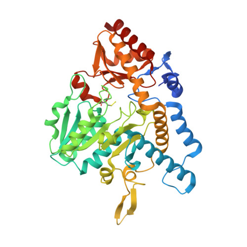
6MRE | pdb_00006mre

E. coli cysteine desulfurase SufS R92A with a cysteine persulfide intermediate


Experimental Data Snapshot

  • Method: X-RAY DIFFRACTION
  • Resolution: 2.50 Å
  • R-Value Free: 
    0.199 (Depositor), 0.200 (DCC) 
  • R-Value Work: 
    0.178 (Depositor), 0.180 (DCC) 
  • R-Value Observed: 
    0.179 (Depositor) 

Starting Model: experimental
View more details

wwPDB Validation   3D Report Full Report


Ligand Structure Quality Assessment 

Created with Raphaël 2.3.0Worse 01 BetterLigand structure goodness of fit to experimental dataBest fitted PLPClick on this verticalbar to view details

This is version 1.4 of the entry. See complete history


Literature

Structural Evidence for Dimer-Interface-Driven Regulation of the Type II Cysteine Desulfurase, SufS.

Dunkle, J.A.Bruno, M.R.Outten, F.W.Frantom, P.A.

(2019) Biochemistry 58: 687-696

  • DOI: https://doi.org/10.1021/acs.biochem.8b01122
  • Primary Citation of Related Structures:  
    6MR2, 6MR6, 6MRE, 6MRH, 6MRI

  • PubMed Abstract: 

    SufS is a type II cysteine desulfurase and acts as the initial step in the Suf Fe-S cluster assembly pathway. In Escherichia coli, this pathway is utilized under conditions of oxidative stress and is resistant to reactive oxygen species. Mechanistically, this means SufS must shift between protecting a covalent persulfide intermediate and making it available for transfer to the next protein partner in the pathway, SufE. Here, we report five X-ray crystal structures of SufS including a new structure of SufS containing an inward-facing persulfide intermediate on C364. Additional structures of SufS variants with substitutions at the dimer interface show changes in dimer geometry and suggest a conserved β-hairpin structure plays a role in mediating interactions with SufE. These new structures, along with previous HDX-MS and biochemical data, identify an interaction network capable of communication between active-sites of the SufS dimer coordinating the shift between desulfurase and transpersulfurase activities.


  • Organizational Affiliation

    Department of Chemistry & Biochemistry , The University of Alabama , Tuscaloosa , Alabama 35487 , United States.


Macromolecules
Find similar proteins by:  (by identity cutoff)  |  3D Structure
Entity ID: 1
MoleculeChains Sequence LengthOrganismDetailsImage
Cysteine desulfurase420Escherichia coli K-12Mutation(s): 1 
Gene Names: sufScsdBynhBb1680JW1670
EC: 2.8.1.7 (PDB Primary Data), 4.4.1.16 (PDB Primary Data), 3.13.1 (UniProt)
UniProt
Find proteins for P77444 (Escherichia coli (strain K12))
Explore P77444 
Go to UniProtKB:  P77444
Entity Groups  
Sequence Clusters30% Identity50% Identity70% Identity90% Identity95% Identity100% Identity
UniProt GroupP77444
Sequence Annotations
Expand
  • Reference Sequence
Small Molecules
Ligands 1 Unique
IDChains Name / Formula / InChI Key2D Diagram3D Interactions
PLP
Query on PLP

Download Ideal Coordinates CCD File 
B [auth A]PYRIDOXAL-5'-PHOSPHATE
C8 H10 N O6 P
NGVDGCNFYWLIFO-UHFFFAOYSA-N
Modified Residues  1 Unique
IDChains TypeFormula2D DiagramParent
CSS
Query on CSS
A
L-PEPTIDE LINKINGC3 H7 N O2 S2CYS
Experimental Data & Validation

Experimental Data

  • Method: X-RAY DIFFRACTION
  • Resolution: 2.50 Å
  • R-Value Free:  0.199 (Depositor), 0.200 (DCC) 
  • R-Value Work:  0.178 (Depositor), 0.180 (DCC) 
  • R-Value Observed: 0.179 (Depositor) 
Space Group: P 43 21 2
Unit Cell:
Length ( Å )Angle ( ˚ )
a = 126.98α = 90
b = 126.98β = 90
c = 135.22γ = 90
Software Package:
Software NamePurpose
XSCALEdata scaling
PHASERphasing
PHENIXrefinement
PDB_EXTRACTdata extraction
XDSdata reduction

Structure Validation

View Full Validation Report


Ligand Structure Quality Assessment 

Created with Raphaël 2.3.0Worse 01 BetterLigand structure goodness of fit to experimental dataBest fitted PLPClick on this verticalbar to view details

Entry History & Funding Information

Deposition Data


Funding OrganizationLocationGrant Number
National Institutes of Health/National Institute of General Medical Sciences (NIH/NIGMS)United StatesGM112919

Revision History  (Full details and data files)

  • Version 1.0: 2019-01-09
    Type: Initial release
  • Version 1.1: 2019-02-27
    Changes: Data collection, Database references
  • Version 1.2: 2020-01-01
    Changes: Author supporting evidence
  • Version 1.3: 2023-10-11
    Changes: Data collection, Database references, Derived calculations, Refinement description
  • Version 1.4: 2023-11-15
    Changes: Data collection