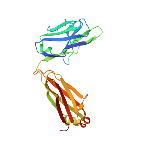
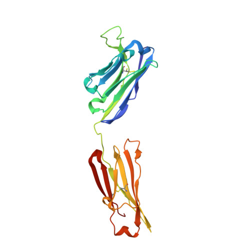
6N32

Anti-HIV-1 Fab 2G12 re-refinement


Experimental Data Snapshot

  • Method: X-RAY DIFFRACTION
  • Resolution: 2.20 Å
  • R-Value Free: 0.227 
  • R-Value Work: 0.198 
  • R-Value Observed: 0.199 

Starting Model: experimental
View more details

wwPDB Validation   3D Report Full Report


This is version 1.3 of the entry. See complete history


Literature

Antibody domain exchange is an immunological solution to carbohydrate cluster recognition.

Calarese, D.A.Scanlan, C.N.Zwick, M.B.Deechongkit, S.Mimura, Y.Kunert, R.Zhu, P.Wormald, M.R.Stanfield, R.L.Roux, K.H.Kelly, J.W.Rudd, P.M.Dwek, R.A.Katinger, H.Burton, D.R.Wilson, I.A.

(2003) Science 300: 2065-2071

  • DOI: https://doi.org/10.1126/science.1083182
  • Primary Citation of Related Structures:  
    6N2X, 6N32, 6N35

  • PubMed Abstract: 

    Human antibody 2G12 neutralizes a broad range of human immunodeficiency virus type 1 (HIV-1) isolates by binding an unusually dense cluster of carbohydrate moieties on the "silent" face of the gp120 envelope glycoprotein. Crystal structures of Fab 2G12 and its complexes with the disaccharide Manalpha1-2Man and with the oligosaccharide Man9GlcNAc2 revealed that two Fabs assemble into an interlocked VH domain-swapped dimer. Further biochemical, biophysical, and mutagenesis data strongly support a Fab-dimerized antibody as the prevalent form that recognizes gp120. The extraordinary configuration of this antibody provides an extended surface, with newly described binding sites, for multivalent interaction with a conserved cluster of oligomannose type sugars on the surface of gp120. The unique interdigitation of Fab domains within an antibody uncovers a previously unappreciated mechanism for high-affinity recognition of carbohydrate or other repeating epitopes on cell or microbial surfaces.


  • Organizational Affiliation

    Department of Molecular Biology, The Scripps Research Institute, 10550 North Torrey Pines Road, La Jolla, CA 92037, USA.


Macromolecules
Find similar proteins by:  (by identity cutoff)  |  3D Structure
Entity ID: 1
MoleculeChains Sequence LengthOrganismDetailsImage
Fab 2G12 light chainA [auth L],
C [auth M]
213Homo sapiensMutation(s): 0 
UniProt
Find proteins for P0DOX7 (Homo sapiens)
Explore P0DOX7 
Go to UniProtKB:  P0DOX7
Entity Groups  
Sequence Clusters30% Identity50% Identity70% Identity90% Identity95% Identity100% Identity
UniProt GroupP0DOX7
Sequence Annotations
Expand
  • Reference Sequence
Find similar proteins by:  (by identity cutoff)  |  3D Structure
Entity ID: 2
MoleculeChains Sequence LengthOrganismDetailsImage
Fab 2G12 heavy chainB [auth H],
D [auth K]
225Homo sapiensMutation(s): 0 
UniProt
Find proteins for P0DOX5 (Homo sapiens)
Explore P0DOX5 
Go to UniProtKB:  P0DOX5
Entity Groups  
Sequence Clusters30% Identity50% Identity70% Identity90% Identity95% Identity100% Identity
UniProt GroupP0DOX5
Sequence Annotations
Expand
  • Reference Sequence
Experimental Data & Validation

Experimental Data

  • Method: X-RAY DIFFRACTION
  • Resolution: 2.20 Å
  • R-Value Free: 0.227 
  • R-Value Work: 0.198 
  • R-Value Observed: 0.199 
  • Space Group: P 21 21 21
Unit Cell:
Length ( Å )Angle ( ˚ )
a = 76.821α = 90
b = 94.182β = 90
c = 171.112γ = 90
Software Package:
Software NamePurpose
PHENIXrefinement
HKL-2000data reduction
SCALEPACKdata scaling
AMoREphasing

Structure Validation

View Full Validation Report


Entry History & Funding Information

Deposition Data


Funding OrganizationLocationGrant Number
National Institutes of Health/National Institute of General Medical Sciences (NIH/NIGMS)United StatesGM46192

Revision History  (Full details and data files)

  • Version 1.0: 2018-11-28
    Type: Initial release
  • Version 1.1: 2020-01-01
    Changes: Author supporting evidence
  • Version 1.2: 2023-10-11
    Changes: Data collection, Database references, Refinement description
  • Version 1.3: 2024-11-20
    Changes: Structure summary