6P3U | pdb_00006p3u

Crystal structure of Eis from Mycobacterium tuberculosis in complex with inhibitor SGT335


Experimental Data Snapshot

  • Method: X-RAY DIFFRACTION
  • Resolution: 2.55 Å
  • R-Value Free: 
    0.206 (Depositor), 0.211 (DCC) 
  • R-Value Work: 
    0.176 (Depositor), 0.181 (DCC) 
  • R-Value Observed: 
    0.177 (Depositor) 

Starting Model: experimental
View more details

wwPDB Validation 3D Report Full Report

Validation slider image for 6P3U

Ligand Structure Quality Assessment 


This is version 1.3 of the entry. See complete history

Literature

Probing the Robustness of Inhibitors of Tuberculosis Aminoglycoside Resistance Enzyme Eis by Mutagenesis.

Green, K.D.Punetha, A.Hou, C.Garneau-Tsodikova, S.Tsodikov, O.V.

(2019) ACS Infect Dis 5: 1772-1778

  • DOI: https://doi.org/10.1021/acsinfecdis.9b00228
  • Primary Citation Related Structures: 
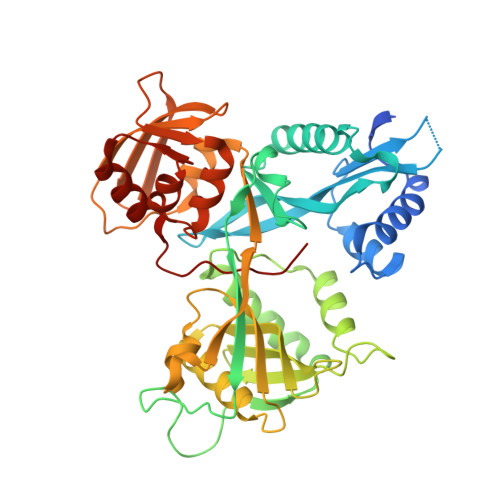
    6P3T, 6P3U, 6P3V

  • PubMed Abstract: 

    Each year, millions of people worldwide contract tuberculosis (TB), the deadliest infection. The spread of infections with drug-resistant strains of Mycobacterium tuberculosis ( Mtb ) that are refractory to treatment poses a major global challenge. A major cause of resistance to antitubercular drugs of last resort, aminoglycosides, is overexpression of the Eis (enhanced intracellular survival) enzyme of Mtb , which inactivates aminoglycosides by acetylating them. We showed previously that this inactivation of aminoglycosides could be overcome by our recently reported Eis inhibitors that are currently in development as potential aminoglycoside adjunctive therapeutics against drug-resistant TB. To interrogate the robustness of the Eis inhibitors, we investigated the enzymatic activity of Eis and its inhibition by Eis inhibitors from three different structural families for nine single-residue mutants of Eis, including those found in the clinic. Three engineered mutations of the substrate binding site, D26A, W36A, and F84A, abolished inhibitor binding while compromising Eis enzymatic activity 2- to 3-fold. All other Eis mutants, including clinically observed ones, were potently inhibited by at least one inhibitor. This study helps position us one step ahead of Mtb resistance to Eis inhibitors as they are being developed for TB therapy.


  • Organizational Affiliation
    • Department of Pharmaceutical Sciences, College of Pharmacy , University of Kentucky , 789 South Limestone Street , Lexington , Kentucky 40536-0596 , United States.

Macromolecule Content 

  • Total Structure Weight: 47.12 kDa 
  • Atom Count: 3,269 
  • Modeled Residue Count: 396 
  • Deposited Residue Count: 422 
  • Unique protein chains: 1

Macromolecules

Find similar proteins by:|  3D Structure
Entity ID: 1
MoleculeChains  Sequence LengthOrganismDetailsImage
N-acetyltransferase Eis422Mycobacterium tuberculosis H37RvMutation(s): 1 
Gene Names: eisRv2416cMTCY253.04
EC: 2.3.1
UniProt
Find proteins for P9WFK7 (Mycobacterium tuberculosis (strain ATCC 25618 / H37Rv))
Explore P9WFK7 
Go to UniProtKB:  P9WFK7
Entity Groups
Sequence Clusters30% Identity50% Identity70% Identity90% Identity95% Identity100% Identity
UniProt GroupP9WFK7
Sequence Annotations
Expand
Reference Sequence

Small Molecules

Ligands 4 Unique
IDChains Name / Formula / InChI Key2D Diagram3D Interactions
NRY
(Subject of Investigation/LOI)

Query on NRY


Download:Ideal Coordinates CCD File
B [auth A]1-(4-fluorophenyl)-2-[2-(4-fluorophenyl)-2-oxoethyl]pyrrolo[1,2-a]pyrazin-2-ium
C21 H15 F2 N2 O
QVAOSDLYVRSWEJ-UHFFFAOYSA-N
PEG

Query on PEG


Download:Ideal Coordinates CCD File
E [auth A]DI(HYDROXYETHYL)ETHER
C4 H10 O3
MTHSVFCYNBDYFN-UHFFFAOYSA-N
SO4

Query on SO4


Download:Ideal Coordinates CCD File
F [auth A],
G [auth A],
H [auth A],
I [auth A],
J [auth A]
SULFATE ION
O4 S
QAOWNCQODCNURD-UHFFFAOYSA-L
GOL

Query on GOL


Download:Ideal Coordinates CCD File
C [auth A],
D [auth A]
GLYCEROL
C3 H8 O3
PEDCQBHIVMGVHV-UHFFFAOYSA-N

Experimental Data & Validation

Experimental Data

  • Method: X-RAY DIFFRACTION
  • Resolution: 2.55 Å
  • R-Value Free:  0.206 (Depositor), 0.211 (DCC) 
  • R-Value Work:  0.176 (Depositor), 0.181 (DCC) 
  • R-Value Observed: 0.177 (Depositor) 
Space Group: H 3 2
Unit Cell:
Length ( Å )Angle ( ˚ )
a = 175.441α = 90
b = 175.441β = 90
c = 122.946γ = 120
Software Package:
Software NamePurpose
REFMACrefinement
HKL-2000data reduction
HKL-2000data scaling
PHASERphasing

Structure Validation

View Full Validation Report


Ligand Structure Quality Assessment 


Entry History 

& Funding Information

Deposition Data


Funding OrganizationLocationGrant Number
National Institutes of Health/National Institute Of Allergy and Infectious Diseases (NIH/NIAID)United StatesR01AI090048

Revision History  (Full details and data files)

  • Version 1.0: 2019-09-04
    Type: Initial release
  • Version 1.1: 2019-10-23
    Changes: Data collection, Database references
  • Version 1.2: 2019-12-18
    Changes: Author supporting evidence
  • Version 1.3: 2023-10-11
    Changes: Data collection, Database references, Refinement description