6QAD | pdb_00006qad

Human Butyrylcholinesterase in complex with ((S)-2-(butylamino)-N-(2-(4-(dimethylamino)cyclohexyl)ethyl)-3-(1H-indol-3-yl)propanamide


Experimental Data Snapshot

  • Method: X-RAY DIFFRACTION
  • Resolution: 2.50 Å
  • R-Value Free: 
    0.234 (Depositor), 0.237 (DCC) 
  • R-Value Work: 
    0.200 (Depositor), 0.201 (DCC) 
  • R-Value Observed: 
    0.202 (Depositor) 

Starting Model: experimental
View more details

wwPDB Validation 3D Report Full Report

Validation slider image for 6QAD

Ligand Structure Quality Assessment 


This is version 2.2 of the entry. See complete history

Literature

Tryptophan-derived butyrylcholinesterase inhibitors as promising leads against Alzheimer's disease.

Meden, A.Knez, D.Jukic, M.Brazzolotto, X.Grsic, M.Pislar, A.Zahirovic, A.Kos, J.Nachon, F.Svete, J.Gobec, S.Groselj, U.

(2019) Chem Commun (Camb) 55: 3765-3768

  • DOI: https://doi.org/10.1039/c9cc01330j
  • Primary Citation Related Structures: 
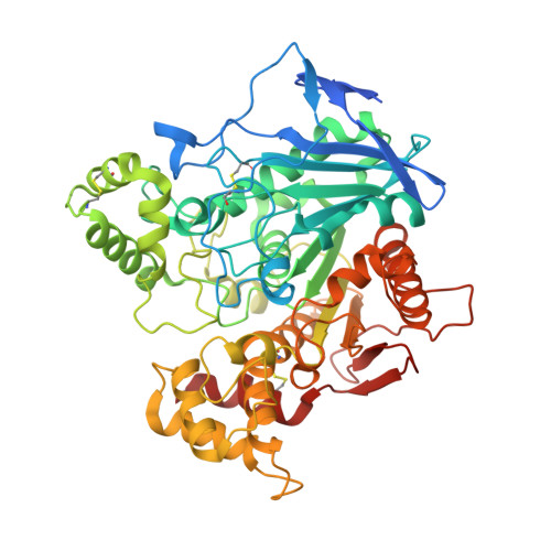
    6QAA, 6QAB, 6QAC, 6QAD, 6QAE

  • PubMed Abstract: 

    We have identified tryptophan-based selective nanomolar butyrylcholinesterase (BChE) inhibitors. They are defined according to their chemical modularity, novel binding mode revealed by five solved crystal structures with human BChE, low cytotoxicity, and predicted permeability of the blood-brain barrier. Altogether, these factors indicate their potential as unique lead compounds for symptomatic therapy against Alzheimer's disease.


  • Organizational Affiliation
    • Faculty of Chemistry and Chemical Technology, University of Ljubljana, Večna pot 113, SI-1000 Ljubljana, Slovenia. uros.groselj@fkkt.uni-lj.si.

Macromolecule Content 

  • Total Structure Weight: 66.64 kDa 
  • Atom Count: 4,530 
  • Modeled Residue Count: 526 
  • Deposited Residue Count: 557 
  • Unique protein chains: 1

Macromolecules

Find similar proteins by:|  3D Structure
Entity ID: 1
MoleculeChains  Sequence LengthOrganismDetailsImage
Cholinesterase557Homo sapiensMutation(s): 4 
Gene Names: BCHECHE1
EC: 3.1.1.8
UniProt & NIH Common Fund Data Resources
Find proteins for P06276 (Homo sapiens)
Explore P06276 
Go to UniProtKB:  P06276
PHAROS:  P06276
GTEx:  ENSG00000114200 
Entity Groups
Sequence Clusters30% Identity50% Identity70% Identity90% Identity95% Identity100% Identity
UniProt GroupP06276
Glycosylation
Glycosylation Sites: 6Go to GlyGen: P06276-1
Sequence Annotations
Expand
Reference Sequence

Oligosaccharides

Help  
Entity ID: 2
MoleculeChains Length2D Diagram GlycosylationD Interactions
alpha-L-fucopyranose-(1-6)-2-acetamido-2-deoxy-beta-D-glucopyranose
B
2N-Glycosylation
Glycosylation Resources
GlyTouCan: G86851RC
GlyCosmos: G86851RC
GlyGen: G86851RC
Entity ID: 3
MoleculeChains Length2D Diagram GlycosylationD Interactions
2-acetamido-2-deoxy-beta-D-glucopyranose-(1-4)-[alpha-L-fucopyranose-(1-6)]2-acetamido-2-deoxy-beta-D-glucopyranose
C, D
3N-Glycosylation
Glycosylation Resources
GlyTouCan: G21290RB
GlyCosmos: G21290RB
GlyGen: G21290RB

Small Molecules

Ligands 6 Unique
IDChains Name / Formula / InChI Key2D Diagram3D Interactions
HUZ
(Subject of Investigation/LOI)

Query on HUZ


Download:Ideal Coordinates CCD File
N [auth A](2~{S})-2-(butylamino)-~{N}-[2-[4-(dimethylamino)cyclohexyl]ethyl]-3-(1~{H}-indol-3-yl)propanamide
C25 H40 N4 O
NDNXTWYOKPDDNQ-PTLVVNQVSA-N
NAG

Query on NAG


Download:Ideal Coordinates CCD File
E [auth A],
F [auth A],
G [auth A]
2-acetamido-2-deoxy-beta-D-glucopyranose
C8 H15 N O6
OVRNDRQMDRJTHS-FMDGEEDCSA-N
MES

Query on MES


Download:Ideal Coordinates CCD File
M [auth A]2-(N-MORPHOLINO)-ETHANESULFONIC ACID
C6 H13 N O4 S
SXGZJKUKBWWHRA-UHFFFAOYSA-N
SO4

Query on SO4


Download:Ideal Coordinates CCD File
O [auth A],
P [auth A],
Q [auth A],
R [auth A]
SULFATE ION
O4 S
QAOWNCQODCNURD-UHFFFAOYSA-L
GOL

Query on GOL


Download:Ideal Coordinates CCD File
I [auth A],
J [auth A],
K [auth A],
L [auth A]
GLYCEROL
C3 H8 O3
PEDCQBHIVMGVHV-UHFFFAOYSA-N
DMS

Query on DMS


Download:Ideal Coordinates CCD File
H [auth A]DIMETHYL SULFOXIDE
C2 H6 O S
IAZDPXIOMUYVGZ-UHFFFAOYSA-N

Experimental Data & Validation

Experimental Data

  • Method: X-RAY DIFFRACTION
  • Resolution: 2.50 Å
  • R-Value Free:  0.234 (Depositor), 0.237 (DCC) 
  • R-Value Work:  0.200 (Depositor), 0.201 (DCC) 
  • R-Value Observed: 0.202 (Depositor) 
Space Group: I 4 2 2
Unit Cell:
Length ( Å )Angle ( ˚ )
a = 155.269α = 90
b = 155.269β = 90
c = 129.922γ = 90
Software Package:
Software NamePurpose
PHENIXrefinement
Cootmodel building
XDSdata reduction
XDSdata scaling
PHENIXphasing

Structure Validation

View Full Validation Report


Ligand Structure Quality Assessment 


Entry History 

& Funding Information

Deposition Data


Funding OrganizationLocationGrant Number
French Ministry of Armed ForcesFrancePDH-2-NRBC-3-C-3201

Revision History  (Full details and data files)

  • Version 1.0: 2019-03-27
    Type: Initial release
  • Version 1.1: 2019-04-03
    Changes: Data collection, Database references
  • Version 2.0: 2020-07-29
    Type: Remediation
    Reason: Carbohydrate remediation
    Changes: Atomic model, Data collection, Derived calculations, Structure summary
  • Version 2.1: 2024-01-24
    Changes: Data collection, Database references, Derived calculations, Refinement description, Structure summary
  • Version 2.2: 2024-11-20
    Changes: Structure summary