6QD5 | pdb_00006qd5

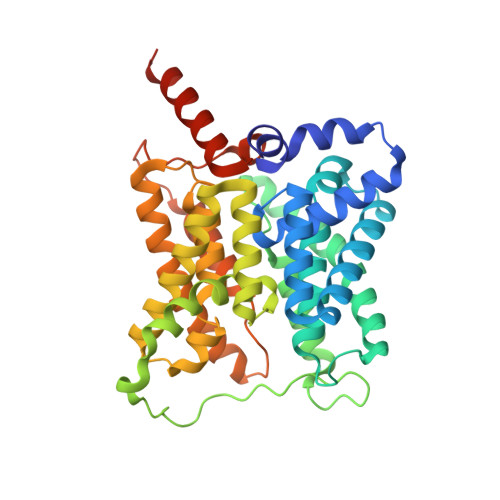
X-ray Structure of the Human Urea Channel SLC14A1/UT1


Experimental Data Snapshot

  • Method: X-RAY DIFFRACTION
  • Resolution: 2.40 Å
  • R-Value Free: 
    0.225 (Depositor), 0.230 (DCC) 
  • R-Value Work: 
    0.175 (Depositor), 0.180 (DCC) 
  • R-Value Observed: 
    0.178 (Depositor) 

Starting Model: experimental
View more details

wwPDB Validation   3D Report Full Report


Ligand Structure Quality Assessment 

Created with Raphaël 2.3.0Worse 01 BetterLigand structure goodness of fit to experimental dataBest fitted Y01Click on this verticalbar to view detailsBest fitted BOGClick on this verticalbar to view details

This is version 1.2 of the entry. See complete history


Macromolecules
Find similar proteins by:  (by identity cutoff)  |  3D Structure
Entity ID: 1
MoleculeChains Sequence LengthOrganismDetailsImage
Urea transporter 1367Homo sapiensMutation(s): 3 
Gene Names: SLC14A1HUT11JKRACH1UT1UTE
Membrane Entity: Yes 
UniProt & NIH Common Fund Data Resources
Find proteins for Q13336 (Homo sapiens)
Explore Q13336 
Go to UniProtKB:  Q13336
PHAROS:  Q13336
GTEx:  ENSG00000141469 
Entity Groups  
Sequence Clusters30% Identity50% Identity70% Identity90% Identity95% Identity100% Identity
UniProt GroupQ13336
Sequence Annotations
Expand
  • Reference Sequence
Small Molecules
Ligands 3 Unique
IDChains Name / Formula / InChI Key2D Diagram3D Interactions
Y01
Query on Y01

Download Ideal Coordinates CCD File 
I [auth A]CHOLESTEROL HEMISUCCINATE
C31 H50 O4
WLNARFZDISHUGS-MIXBDBMTSA-N
BOG
Query on BOG

Download Ideal Coordinates CCD File 
B [auth A]
C [auth A]
D [auth A]
E [auth A]
F [auth A]
octyl beta-D-glucopyranoside
C14 H28 O6
HEGSGKPQLMEBJL-RKQHYHRCSA-N
PG4
Query on PG4

Download Ideal Coordinates CCD File 
J [auth A],
K [auth A]
TETRAETHYLENE GLYCOL
C8 H18 O5
UWHCKJMYHZGTIT-UHFFFAOYSA-N
Experimental Data & Validation

Experimental Data

  • Method: X-RAY DIFFRACTION
  • Resolution: 2.40 Å
  • R-Value Free:  0.225 (Depositor), 0.230 (DCC) 
  • R-Value Work:  0.175 (Depositor), 0.180 (DCC) 
  • R-Value Observed: 0.178 (Depositor) 
Space Group: P 63
Unit Cell:
Length ( Å )Angle ( ˚ )
a = 112.331α = 90
b = 112.331β = 90
c = 87.94γ = 120
Software Package:
Software NamePurpose
PHENIXrefinement
XDSdata reduction
PHENIXphasing

Structure Validation

View Full Validation Report


Ligand Structure Quality Assessment 

Created with Raphaël 2.3.0Worse 01 BetterLigand structure goodness of fit to experimental dataBest fitted Y01Click on this verticalbar to view detailsBest fitted BOGClick on this verticalbar to view details

Entry History & Funding Information

Revision History  (Full details and data files)

  • Version 1.0: 2019-01-09
    Type: Initial release
  • Version 1.1: 2020-07-29
    Type: Remediation
    Reason: Carbohydrate remediation
    Changes: Data collection, Derived calculations, Structure summary
  • Version 1.2: 2024-01-24
    Changes: Advisory, Data collection, Database references, Refinement description, Structure summary