6RJ4 | pdb_00006rj4

Molybdenum storage protein - P6422, ADP


Experimental Data Snapshot

  • Method: X-RAY DIFFRACTION
  • Resolution: 1.90 Å
  • R-Value Free: 
    0.200 (Depositor), 0.201 (DCC) 
  • R-Value Work: 
    0.172 (Depositor), 0.173 (DCC) 
  • R-Value Observed: 
    0.173 (Depositor) 

Starting Model: experimental
View more details

wwPDB Validation 3D Report Full Report

Validation slider image for 6RJ4

Ligand Structure Quality Assessment 


This is version 1.1 of the entry. See complete history

Literature

Molybdate pumping into the molybdenum storage protein via an ATP-powered piercing mechanism.

Brunle, S.Eisinger, M.L.Poppe, J.Mills, D.J.Langer, J.D.Vonck, J.Ermler, U.

(2019) Proc Natl Acad Sci U S A 

  • DOI: https://doi.org/10.1073/pnas.1913031116
  • Primary Citation Related Structures: 
    6RIS, 6RJ4, 6RKD, 6RKE

  • PubMed Abstract: 

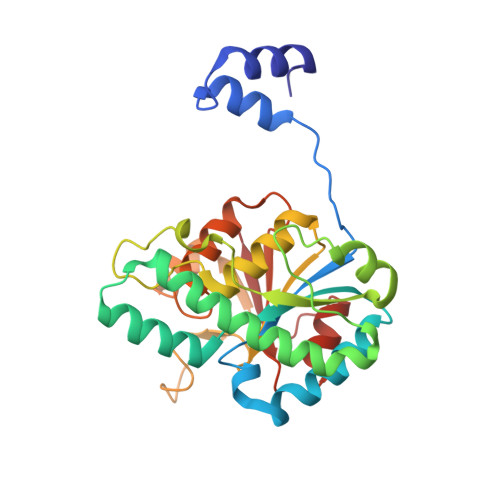
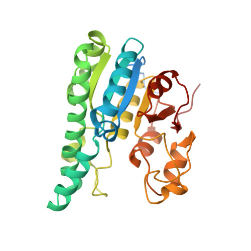
    The molybdenum storage protein (MoSto) deposits large amounts of molybdenum as polyoxomolybdate clusters in a heterohexameric (αβ) 3 cage-like protein complex under ATP consumption. Here, we suggest a unique mechanism for the ATP-powered molybdate pumping process based on X-ray crystallography, cryoelectron microscopy, hydrogen-deuterium exchange mass spectrometry, and mutational studies of MoSto from Azotobacter vinelandii . First, we show that molybdate, ATP, and Mg 2+ consecutively bind into the open ATP-binding groove of the β-subunit, which thereafter becomes tightly locked by fixing the previously disordered N-terminal arm of the α-subunit over the β-ATP. Next, we propose a nucleophilic attack of molybdate onto the γ-phosphate of β-ATP, analogous to the similar reaction of the structurally related UMP kinase. The formed instable phosphoric-molybdic anhydride becomes immediately hydrolyzed and, according to the current data, the released and accelerated molybdate is pressed through the cage wall, presumably by turning aside the Metβ149 side chain. A structural comparison between MoSto and UMP kinase provides valuable insight into how an enzyme is converted into a molecular machine during evolution. The postulated direct conversion of chemical energy into kinetic energy via an activating molybdate kinase and an exothermic pyrophosphatase reaction to overcome a proteinous barrier represents a novelty in ATP-fueled biochemistry, because normally, ATP hydrolysis initiates large-scale conformational changes to drive a distant process.


  • Organizational Affiliation
    • Department of Molecular Membrane Biology, Max Planck Institute of Biophysics, 60438 Frankfurt am Main, Germany.

Macromolecule Content 

  • Total Structure Weight: 175.97 kDa 
  • Atom Count: 12,665 
  • Modeled Residue Count: 1,550 
  • Deposited Residue Count: 1,632 
  • Unique protein chains: 2

Macromolecules

Find similar proteins by:|  3D Structure
Entity ID: 1
MoleculeChains  Sequence LengthOrganismDetailsImage
Molybdenum storage protein subunit betaA [auth B],
C [auth D],
E [auth F]
269Azotobacter vinelandii DJMutation(s): 0 
Gene Names: mosBAvin_43210
UniProt
Find proteins for P84253 (Azotobacter vinelandii (strain DJ / ATCC BAA-1303))
Explore P84253 
Go to UniProtKB:  P84253
Entity Groups
Sequence Clusters30% Identity50% Identity70% Identity90% Identity95% Identity100% Identity
UniProt GroupP84253
Sequence Annotations
Expand
Reference Sequence
Find similar proteins by:|  3D Structure
Entity ID: 2
MoleculeChains  Sequence LengthOrganismDetailsImage
Molybdenum storage protein subunit alphaB [auth A],
D [auth C],
F [auth E]
275Azotobacter vinelandii DJMutation(s): 0 
Gene Names: mosAAvin_43200
UniProt
Find proteins for P84308 (Azotobacter vinelandii (strain DJ / ATCC BAA-1303))
Explore P84308 
Go to UniProtKB:  P84308
Entity Groups
Sequence Clusters30% Identity50% Identity70% Identity90% Identity95% Identity100% Identity
UniProt GroupP84308
Sequence Annotations
Expand
Reference Sequence

Small Molecules

Ligands 5 Unique
IDChains Name / Formula / InChI Key2D Diagram3D Interactions
ATP

Query on ATP


Download:Ideal Coordinates CCD File
L [auth A],
Q [auth C],
V [auth E]
ADENOSINE-5'-TRIPHOSPHATE
C10 H16 N5 O13 P3
ZKHQWZAMYRWXGA-KQYNXXCUSA-N
ADP
(Subject of Investigation/LOI)

Query on ADP


Download:Ideal Coordinates CCD File
G [auth B],
N [auth D],
S [auth F]
ADENOSINE-5'-DIPHOSPHATE
C10 H15 N5 O10 P2
XTWYTFMLZFPYCI-KQYNXXCUSA-N
PO4

Query on PO4


Download:Ideal Coordinates CCD File
H [auth B]
I [auth B]
J [auth B]
P [auth D]
T [auth F]
H [auth B],
I [auth B],
J [auth B],
P [auth D],
T [auth F],
U [auth F]
PHOSPHATE ION
O4 P
NBIIXXVUZAFLBC-UHFFFAOYSA-K
MG

Query on MG


Download:Ideal Coordinates CCD File
M [auth A],
O [auth D],
R [auth C],
W [auth E]
MAGNESIUM ION
Mg
JLVVSXFLKOJNIY-UHFFFAOYSA-N
NA

Query on NA


Download:Ideal Coordinates CCD File
K [auth B]SODIUM ION
Na
FKNQFGJONOIPTF-UHFFFAOYSA-N

Experimental Data & Validation

Experimental Data

  • Method: X-RAY DIFFRACTION
  • Resolution: 1.90 Å
  • R-Value Free:  0.200 (Depositor), 0.201 (DCC) 
  • R-Value Work:  0.172 (Depositor), 0.173 (DCC) 
  • R-Value Observed: 0.173 (Depositor) 
Space Group: P 64 2 2
Unit Cell:
Length ( Å )Angle ( ˚ )
a = 188.68α = 90
b = 188.68β = 90
c = 188.63γ = 120
Software Package:
Software NamePurpose
PHENIXrefinement
XDSdata reduction
XSCALEdata scaling
PHASERphasing

Structure Validation

View Full Validation Report


Ligand Structure Quality Assessment 


Entry History 

Deposition Data

Revision History  (Full details and data files)

  • Version 1.0: 2019-12-18
    Type: Initial release
  • Version 1.1: 2024-01-24
    Changes: Data collection, Database references, Derived calculations, Refinement description