6RWV | pdb_00006rwv

Structure of apo-LmCpfC


Experimental Data Snapshot

  • Method: X-RAY DIFFRACTION
  • Resolution: 1.64 Å
  • R-Value Free: 
    0.201 (Depositor), 0.203 (DCC) 
  • R-Value Work: 
    0.176 (Depositor), 0.178 (DCC) 
  • R-Value Observed: 
    0.177 (Depositor) 

Starting Model: experimental
View more details

wwPDB Validation 3D Report Full Report

Validation slider image for 6RWV

This is version 1.2 of the entry. See complete history

Literature

Crystal structures and calorimetry reveal catalytically relevant binding mode of coproporphyrin and coproheme in coproporphyrin ferrochelatase.

Hofbauer, S.Helm, J.Obinger, C.Djinovic-Carugo, K.Furtmuller, P.G.

(2020) FEBS J 287: 2779-2796

  • DOI: https://doi.org/10.1111/febs.15164
  • Primary Citation Related Structures: 
    6RWV, 6SV3

  • PubMed Abstract: 

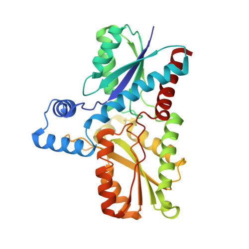
    Coproporphyrin ferrochelatases (CpfCs, EC 4.99.1.9) insert ferrous iron into coproporphyrin III yielding coproheme. CpfCs are utilized by prokaryotic, mainly monoderm (Gram-positive) bacteria within the recently detected coproporphyrin-dependent (CPD) heme biosynthesis pathway. Here, we present a comprehensive study on CpfC from Listeria monocytogenes (LmCpfC) including the first crystal structure of a coproheme-bound CpfC. Comparison of crystal structures of apo-LmCpfC and coproheme-LmCpfC allowed identification of structural rearrangements and of amino acids involved in tetrapyrrole macrocycle and Fe 2+ binding. Differential scanning calorimetry of apo-, coproporphyrin III-, and coproheme-LmCpfC underline the pronounced noncovalent interaction of both coproporphyrin and coproheme with the protein (ΔT m  = 11 °C compared to apo-LmCpfC), which includes the propionates (p2, p4, p6, p7) and the amino acids Arg29, Arg45, Tyr46, Ser53, and Tyr124. Furthermore, the thermodynamics and kinetics of coproporphyrin III and coproheme binding to apo-LmCpfC is presented as well as the kinetics of insertion of ferrous iron into coproporphyrin III-LmCpfC that immediately leads to formation of ferric coproheme-LmCpfC (k cat /K M  = 4.7 × 10 5  m -1 ·s -1 ). We compare the crystal structure of coproheme-LmCpfC with available structures of CpfCs with artificial tetrapyrrole macrocycles and discuss our data on substrate binding, iron insertion and substrate release in the context of the CPD heme biosynthesis pathway. ENZYME: EC 4.99.1.9 DATABASE: pdb-codes of structural data in this work: 6RWV, 6SV3.


  • Organizational Affiliation
    • Department of Chemistry, Institute of Biochemistry, BOKU - University of Natural Resources and Life Sciences, Vienna, Austria.

Macromolecule Content 

  • Total Structure Weight: 36.68 kDa 
  • Atom Count: 2,887 
  • Modeled Residue Count: 310 
  • Deposited Residue Count: 312 
  • Unique protein chains: 1

Macromolecules

Find similar proteins by:|  3D Structure
Entity ID: 1
MoleculeChains  Sequence LengthOrganismDetailsImage
Ferrochelatase312Listeria monocytogenesMutation(s): 0 
Gene Names: 
EC: 4.99.1.1 (PDB Primary Data), 4.99.1.9 (UniProt)
UniProt
Find proteins for Q8Y565 (Listeria monocytogenes serovar 1/2a (strain ATCC BAA-679 / EGD-e))
Explore Q8Y565 
Go to UniProtKB:  Q8Y565
Entity Groups
Sequence Clusters30% Identity50% Identity70% Identity90% Identity95% Identity100% Identity
UniProt GroupQ8Y565
Sequence Annotations
Expand
Reference Sequence

Small Molecules

Ligands 3 Unique
IDChains Name / Formula / InChI Key2D Diagram3D Interactions
PO4

Query on PO4


Download:Ideal Coordinates CCD File
I [auth A],
J [auth A]
PHOSPHATE ION
O4 P
NBIIXXVUZAFLBC-UHFFFAOYSA-K
GOL

Query on GOL


Download:Ideal Coordinates CCD File
B [auth A]
C [auth A]
D [auth A]
E [auth A]
F [auth A]
B [auth A],
C [auth A],
D [auth A],
E [auth A],
F [auth A],
G [auth A],
H [auth A]
GLYCEROL
C3 H8 O3
PEDCQBHIVMGVHV-UHFFFAOYSA-N
K

Query on K


Download:Ideal Coordinates CCD File
K [auth A],
L [auth A]
POTASSIUM ION
K
NPYPAHLBTDXSSS-UHFFFAOYSA-N

Experimental Data & Validation

Experimental Data

  • Method: X-RAY DIFFRACTION
  • Resolution: 1.64 Å
  • R-Value Free:  0.201 (Depositor), 0.203 (DCC) 
  • R-Value Work:  0.176 (Depositor), 0.178 (DCC) 
  • R-Value Observed: 0.177 (Depositor) 
Space Group: P 1 21 1
Unit Cell:
Length ( Å )Angle ( ˚ )
a = 48.303α = 90
b = 76.723β = 106.558
c = 52.258γ = 90
Software Package:
Software NamePurpose
PHENIXrefinement
PHENIXrefinement
XDSdata reduction
XDSdata scaling
PHASERphasing

Structure Validation

View Full Validation Report


Entry History 

& Funding Information

Deposition Data


Funding OrganizationLocationGrant Number
Austrian Science FundAustriaP29099

Revision History  (Full details and data files)

  • Version 1.0: 2019-12-18
    Type: Initial release
  • Version 1.1: 2020-07-15
    Changes: Database references
  • Version 1.2: 2024-01-24
    Changes: Data collection, Database references, Refinement description