6SQC | pdb_00006sqc

Crystal structure of complex between nuclear coactivator binding domain of CBP and [1040-1086]ACTR containing alpha-methylated Leu1055 and Leu1076


Experimental Data Snapshot

  • Method: X-RAY DIFFRACTION
  • Resolution: 2.28 Å
  • R-Value Free: 
    0.274 (Depositor), 0.276 (DCC) 
  • R-Value Work: 
    0.228 (Depositor), 0.229 (DCC) 
  • R-Value Observed: 
    0.230 (Depositor) 

Starting Model: experimental
View more details

wwPDB Validation 3D Report Full Report

Validation slider image for 6SQC

This is version 1.4 of the entry. See complete history

Literature

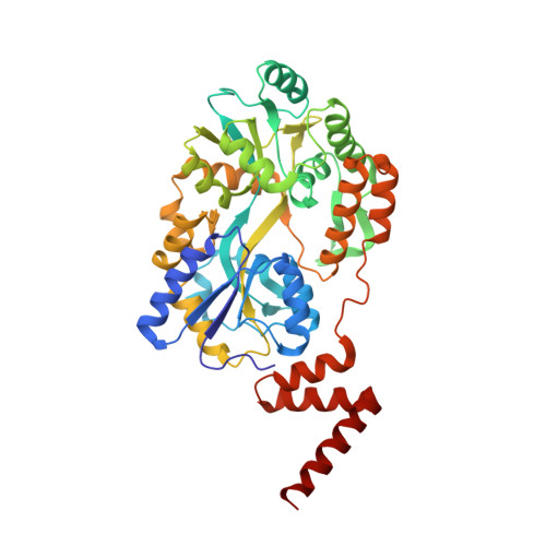
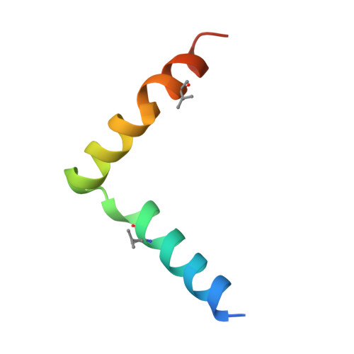
Conformational editing of intrinsically disordered protein by alpha-methylation.

Bauer, V.Schmidtgall, B.Gogl, G.Dolenc, J.Osz, J.Nomine, Y.Kostmann, C.Cousido-Siah, A.Mitschler, A.Rochel, N.Trave, G.Kieffer, B.Torbeev, V.

(2020) Chem Sci 12: 1080-1089

  • DOI: https://doi.org/10.1039/d0sc04482b
  • Primary Citation Related Structures: 
    6SQC

  • PubMed Abstract: 

    Intrinsically disordered proteins (IDPs) constitute a large portion of "Dark Proteome" - difficult to characterize or yet to be discovered protein structures. Here we used conformationally constrained α-methylated amino acids to bias the conformational ensemble in the free unstructured activation domain of transcriptional coactivator ACTR. Different sites and patterns of substitutions were enabled by chemical protein synthesis and led to distinct populations of α-helices. A specific substitution pattern resulted in a substantially higher binding affinity to nuclear coactivator binding domain (NCBD) of CREB-binding protein, a natural binding partner of ACTR. The first X-ray structure of the modified ACTR domain - NCBD complex visualized a unique conformation of ACTR and confirmed that the key α-methylated amino acids are localized within α-helices in the bound state. This study demonstrates a strategy for characterization of individual conformational states of IDPs.


  • Organizational Affiliation
    • Institut de Science et d'Ingénierie Supramoléculaires (ISIS), International Center for Frontier Research in Chemistry (icFRC), University of Strasbourg, CNRS, UMR 7006 Strasbourg France torbeev@unistra.fr.

Macromolecule Content 

  • Total Structure Weight: 51.96 kDa 
  • Atom Count: 3,759 
  • Modeled Residue Count: 465 
  • Deposited Residue Count: 471 
  • Unique protein chains: 2

Macromolecules

Find similar proteins by:|  3D Structure
Entity ID: 1
MoleculeChains  Sequence LengthOrganismDetailsImage
Maltose/maltodextrin-binding periplasmic protein,CREB-binding protein424Escherichia coli K-12Homo sapiens
This entity is chimeric
Mutation(s): 0 
Gene Names: malEb4034JW3994CREBBPCBP
EC: 2.3.1.48 (PDB Primary Data), 2.3.1 (UniProt)
UniProt & NIH Common Fund Data Resources
Find proteins for P0AEX9 (Escherichia coli (strain K12))
Explore P0AEX9 
Go to UniProtKB:  P0AEX9
Find proteins for Q92793 (Homo sapiens)
Explore Q92793 
Go to UniProtKB:  Q92793
PHAROS:  Q92793
GTEx:  ENSG00000005339 
Entity Groups
Sequence Clusters30% Identity50% Identity70% Identity90% Identity95% Identity100% Identity
UniProt GroupsQ92793P0AEX9
Sequence Annotations
Expand
Reference Sequence
Find similar proteins by:|  3D Structure
Entity ID: 2
MoleculeChains  Sequence LengthOrganismDetailsImage
Nuclear receptor coactivator 347Homo sapiensMutation(s): 0 
EC: 2.3.1.48
UniProt & NIH Common Fund Data Resources
Find proteins for Q9Y6Q9 (Homo sapiens)
Explore Q9Y6Q9 
Go to UniProtKB:  Q9Y6Q9
PHAROS:  Q9Y6Q9
GTEx:  ENSG00000124151 
Entity Groups
Sequence Clusters30% Identity50% Identity70% Identity90% Identity95% Identity100% Identity
UniProt GroupQ9Y6Q9
Sequence Annotations
Expand
Reference Sequence

Oligosaccharides

Help  
Entity ID: 3
MoleculeChains Length2D Diagram GlycosylationD Interactions
alpha-D-glucopyranose-(1-4)-alpha-D-glucopyranose
C
2N/A
Glycosylation Resources
GlyTouCan: G07411ON
GlyCosmos: G07411ON

Biologically Interesting Molecules (External Reference) 

1 Unique

Experimental Data & Validation

Experimental Data

  • Method: X-RAY DIFFRACTION
  • Resolution: 2.28 Å
  • R-Value Free:  0.274 (Depositor), 0.276 (DCC) 
  • R-Value Work:  0.228 (Depositor), 0.229 (DCC) 
  • R-Value Observed: 0.230 (Depositor) 
Space Group: C 1 2 1
Unit Cell:
Length ( Å )Angle ( ˚ )
a = 103.18α = 90
b = 42.46β = 101.125
c = 113.79γ = 90
Software Package:
Software NamePurpose
PHENIXrefinement
XDSdata reduction
XSCALEdata scaling
PHASERphasing

Structure Validation

View Full Validation Report


Entry History 

& Funding Information

Deposition Data


Funding OrganizationLocationGrant Number
European Research Council (ERC)France715062-HiChemSynPro
Laboratories of Excellence (LabEx)FranceANR-10-LABX-0026_CSC
ATIP-AvenirFrance--

Revision History  (Full details and data files)

  • Version 1.0: 2020-09-30
    Type: Initial release
  • Version 1.1: 2022-04-20
    Changes: Database references
  • Version 1.2: 2022-12-14
    Changes: Author supporting evidence
  • Version 1.3: 2024-01-31
    Changes: Data collection, Refinement description
  • Version 1.4: 2024-10-16
    Changes: Structure summary