Structural Recognition of Spectinomycin by Resistance Enzyme ANT(9) from Enterococcus faecalis.
Kanchugal P, S., Selmer, M.(2020) Antimicrob Agents Chemother 64
- PubMed: 32253216
- DOI: https://doi.org/10.1128/AAC.00371-20
- Primary Citation of Related Structures:
6SXJ, 6XXQ, 6XZ0 - PubMed Abstract:
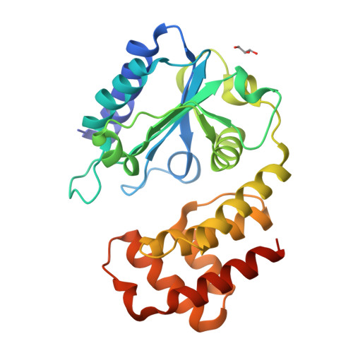
Spectinomycin is a ribosome-binding antibiotic that blocks the translocation step of translation. A prevalent resistance mechanism is modification of the drug by aminoglycoside nucleotidyl transferase (ANT) enzymes of the spectinomycin-specific ANT(9) family or by enzymes of the dual-specificity ANT(3")(9) family, which also acts on streptomycin. We previously reported the structural mechanism of streptomycin modification by the ANT(3")(9) AadA from Salmonella enterica ANT(9) from Enterococcus faecalis adenylates the 9-hydroxyl of spectinomycin. Here, we present the first structures of spectinomycin bound to an ANT enzyme. Structures were solved for ANT(9) in apo form, in complex with ATP, spectinomycin, and magnesium, or in complex with only spectinomycin. ANT(9) shows an overall structure similar to that of AadA, with an N-terminal nucleotidyltransferase domain and a C-terminal α-helical domain. Spectinomycin binds close to the entrance of the interdomain cleft, while ATP is buried at the bottom. Upon drug binding, the C-terminal domain rotates 14 degrees to close the cleft, allowing contacts of both domains with the drug. Comparison with AadA shows that spectinomycin specificity is explained by a straight α 5 helix and a shorter α 5 -α 6 loop, which would clash with the larger streptomycin substrate. In the active site, we observed two magnesium ions, one of them in a previously unobserved position that may activate the 9-hydroxyl for deprotonation by the catalytic base Glu-86. The observed binding mode for spectinomycin suggests that spectinamides and aminomethyl spectinomycins, recent spectinomycin analogues with expansions in position 4 of the C ring, are also subjected to modification by ANT(9) and ANT(3")(9) enzymes.
Organizational Affiliation:
Department of Cell and Molecular Biology, Uppsala University, Uppsala, Sweden.


