6TFL | pdb_00006tfl

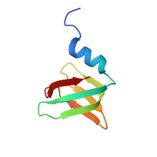
Lsm protein (SmAP) from Halobacterium salinarum


Experimental Data Snapshot

  • Method: X-RAY DIFFRACTION
  • Resolution: 2.40 Å
  • R-Value Free: 
    0.231 (Depositor), 0.233 (DCC) 
  • R-Value Work: 
    0.178 (Depositor), 0.185 (DCC) 
  • R-Value Observed: 
    0.180 (Depositor) 

Starting Model: experimental
View more details

wwPDB Validation 3D Report Full Report

Validation slider image for 6TFL

Ligand Structure Quality Assessment 


This is version 1.2 of the entry. See complete history

Literature

Structure and RNA-Binding Properties of Lsm Protein from Halobacterium salinarum.

Fando, M.S.Mikhaylina, A.O.Lekontseva, N.V.Tishchenko, S.V.Nikulin, A.D.

(2021) Biochemistry (Mosc) 86: 833-842

  • DOI: https://doi.org/10.1134/S000629792107004X
  • Primary Citation Related Structures: 
    6TFL

  • PubMed Abstract: 

    The structure and the RNA-binding properties of the Lsm protein from Halobacterium salinarum have been determined. A distinctive feature of this protein is the presence of a short L4 loop connecting the β3 and β4 strands. Since bacterial Lsm proteins (also called Hfq proteins) have a short L4 loop and form hexamers, whereas archaeal Lsm proteins (SmAP) have a long L4 loop and form heptamers, it has been suggested that the length of the L4 loop may affect the quaternary structure of Lsm proteins. Moreover, the L4 loop covers the region of SmAP corresponding to one of the RNA-binding sites in Hfq, and thus can affect the RNA-binding properties of the protein. Our results show that the SmAP from H. salinarum forms heptamers and possesses the same RNA-binding properties as homologous proteins with the long L4 loop. Therefore, the length of the L4 does not govern the number of monomers in the protein particles and does not affect the RNA-binding properties of Lsm proteins.


  • Organizational Affiliation
    • Institute of Protein Research Russian Academy of Sciences, Pushchino, Moscow Region, 142290, Russia.

Macromolecule Content 

  • Total Structure Weight: 99.38 kDa 
  • Atom Count: 7,184 
  • Modeled Residue Count: 868 
  • Deposited Residue Count: 868 
  • Unique protein chains: 1

Macromolecules

Find similar proteins by:|  3D Structure
Entity ID: 1
MoleculeChains  Sequence LengthOrganismDetailsImage
RNA-binding protein Lsm
A, B, C, D, E
A, B, C, D, E, F, G, H, I, J, K, L, M, N
62Halobacterium salinarum R1Mutation(s): 0 
Gene Names: lsmsnpOE_3142R
UniProt
Find proteins for B0R5R2 (Halobacterium salinarum (strain ATCC 29341 / DSM 671 / R1))
Explore B0R5R2 
Go to UniProtKB:  B0R5R2
Entity Groups
Sequence Clusters30% Identity50% Identity70% Identity90% Identity95% Identity100% Identity
UniProt GroupB0R5R2
Sequence Annotations
Expand
Reference Sequence

Small Molecules

Ligands 3 Unique
IDChains Name / Formula / InChI Key2D Diagram3D Interactions
URI

Query on URI


Download:Ideal Coordinates CCD File
CA [auth E]
EA [auth F]
EB [auth N]
GA [auth G]
KA [auth H]
CA [auth E],
EA [auth F],
EB [auth N],
GA [auth G],
KA [auth H],
OA [auth I],
Q [auth A],
QA [auth J],
T [auth B],
TA [auth K],
U [auth C],
XA [auth L],
Z [auth D],
ZA [auth M]
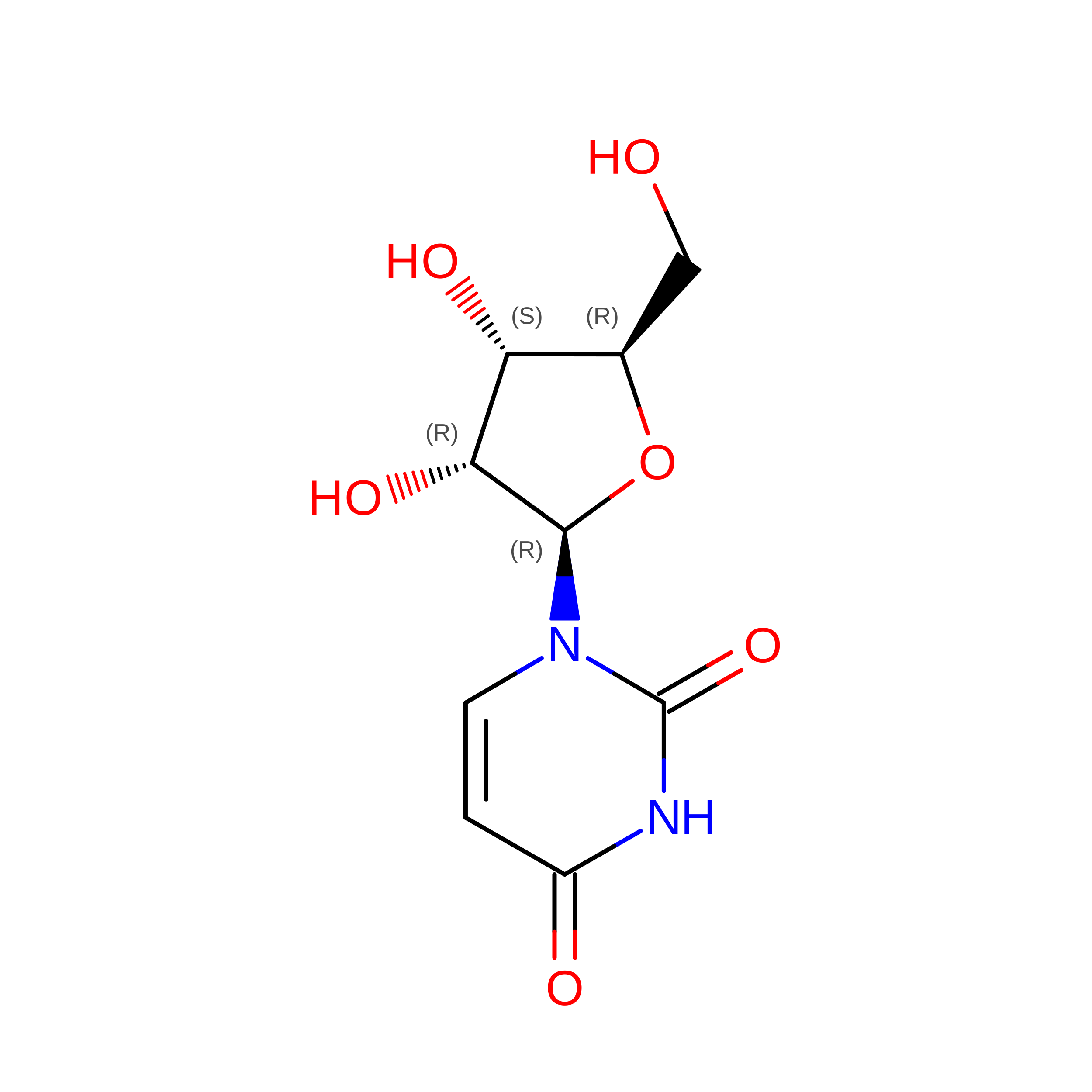
URIDINE
C9 H12 N2 O6
DRTQHJPVMGBUCF-XVFCMESISA-N
GOL

Query on GOL


Download:Ideal Coordinates CCD File
FA [auth G]GLYCEROL
C3 H8 O3
PEDCQBHIVMGVHV-UHFFFAOYSA-N
CA

Query on CA


Download:Ideal Coordinates CCD File
AA [auth E]
AB [auth N]
BA [auth E]
BB [auth N]
CB [auth N]
AA [auth E],
AB [auth N],
BA [auth E],
BB [auth N],
CB [auth N],
DA [auth F],
DB [auth N],
HA [auth H],
IA [auth H],
JA [auth H],
LA [auth I],
MA [auth I],
NA [auth I],
O [auth A],
P [auth A],
PA [auth J],
R [auth B],
RA [auth K],
S [auth B],
SA [auth K],
UA [auth L],
V [auth D],
VA [auth L],
W [auth D],
WA [auth L],
X [auth D],
Y [auth D],
YA [auth M]
CALCIUM ION
Ca
BHPQYMZQTOCNFJ-UHFFFAOYSA-N

Experimental Data & Validation

Experimental Data

  • Method: X-RAY DIFFRACTION
  • Resolution: 2.40 Å
  • R-Value Free:  0.231 (Depositor), 0.233 (DCC) 
  • R-Value Work:  0.178 (Depositor), 0.185 (DCC) 
  • R-Value Observed: 0.180 (Depositor) 
Space Group: P 41 21 2
Unit Cell:
Length ( Å )Angle ( ˚ )
a = 156.45α = 90
b = 156.45β = 90
c = 106.63γ = 90
Software Package:
Software NamePurpose
PHENIXrefinement
PDB_EXTRACTdata extraction
XDSdata reduction
Aimlessdata scaling
MoRDaphasing

Structure Validation

View Full Validation Report


Ligand Structure Quality Assessment 


Entry History 

& Funding Information

Deposition Data


Funding OrganizationLocationGrant Number
Russian Foundation for Basic ResearchRussian Federation18-04-00222

Revision History  (Full details and data files)

  • Version 1.0: 2020-11-25
    Type: Initial release
  • Version 1.1: 2021-12-08
    Changes: Database references
  • Version 1.2: 2024-01-24
    Changes: Data collection, Refinement description