


The structure of penicillin-binding protein 3 from Yersinia pestis
Pankov, G.To be published.
Experimental Data Snapshot
Starting Model: experimental
View more details
wwPDB Validation 3D Report Full Report

Entity ID: 1 | |||||
|---|---|---|---|---|---|
| Molecule | Chains | Sequence Length | Organism | Details | Image |
| Peptidoglycan D,D-transpeptidase FtsI | A [auth AAA], B [auth BBB] | 549 | Yersinia pestis | Mutation(s): 0 Gene Names: ftsI, YPO0549 EC: 3.4.16.4 | 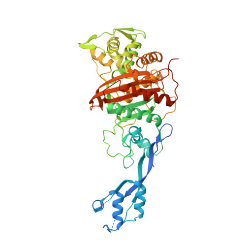 |
UniProt | |||||
Entity Groups | |||||
| Sequence Clusters | 30% Identity50% Identity70% Identity90% Identity95% Identity100% Identity | ||||
| UniProt Group | A0A3N4B5A3 | ||||
Sequence AnnotationsExpand | |||||
Reference Sequence | |||||
| Length ( Å ) | Angle ( ˚ ) |
|---|---|
| a = 100.8 | α = 90 |
| b = 100.8 | β = 90 |
| c = 314.409 | γ = 90 |
| Software Name | Purpose |
|---|---|
| REFMAC | refinement |
| XDS | data reduction |
| Aimless | data scaling |
| PHASER | phasing |


