6TZ6 | pdb_00006tz6

Crystal Structure of Candida Albicans Calcineurin A, Calcineurin B, FKBP12 and FK506 (Tacrolimus)


Experimental Data Snapshot

  • Method: X-RAY DIFFRACTION
  • Resolution: 2.55 Å
  • R-Value Free: 
    0.229 (Depositor), 0.231 (DCC) 
  • R-Value Work: 
    0.182 (Depositor), 0.185 (DCC) 
  • R-Value Observed: 
    0.184 (Depositor) 

Starting Model: experimental
View more details

wwPDB Validation 3D Report Full Report

Validation slider image for 6TZ6

Ligand Structure Quality Assessment 


This is version 1.2 of the entry. See complete history

Literature

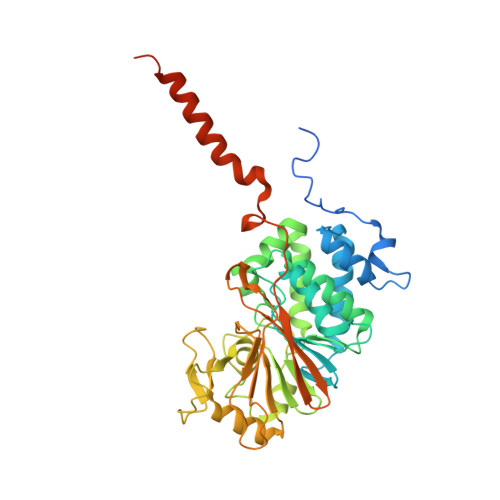
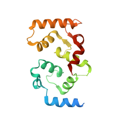
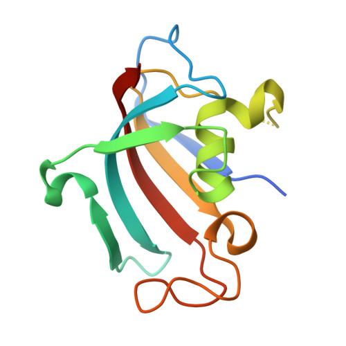
Harnessing calcineurin-FK506-FKBP12 crystal structures from invasive fungal pathogens to develop antifungal agents.

Juvvadi, P.R.Fox 3rd, D.Bobay, B.G.Hoy, M.J.Gobeil, S.M.C.Venters, R.A.Chang, Z.Lin, J.J.Averette, A.F.Cole, D.C.Barrington, B.C.Wheaton, J.D.Ciofani, M.Trzoss, M.Li, X.Lee, S.C.Chen, Y.L.Mutz, M.Spicer, L.D.Schumacher, M.A.Heitman, J.Steinbach, W.J.

(2019) Nat Commun 10: 4275-4275

  • DOI: https://doi.org/10.1038/s41467-019-12199-1
  • Primary Citation Related Structures: 
    5B8I, 6TZ6, 6TZ7, 6TZ8

  • PubMed Abstract: 

    Calcineurin is important for fungal virulence and a potential antifungal target, but compounds targeting calcineurin, such as FK506, are immunosuppressive. Here we report the crystal structures of calcineurin catalytic (CnA) and regulatory (CnB) subunits complexed with FK506 and the FK506-binding protein (FKBP12) from human fungal pathogens (Aspergillus fumigatus, Candida albicans, Cryptococcus neoformans and Coccidioides immitis). Fungal calcineurin complexes are similar to the mammalian complex, but comparison of fungal and human FKBP12 (hFKBP12) reveals conformational differences in the 40s and 80s loops. NMR analysis, molecular dynamic simulations, and mutations of the A. fumigatus CnA/CnB-FK506-FKBP12-complex identify a Phe88 residue, not conserved in hFKBP12, as critical for binding and inhibition of fungal calcineurin. These differences enable us to develop a less immunosuppressive FK506 analog, APX879, with an acetohydrazine substitution of the C22-carbonyl of FK506. APX879 exhibits reduced immunosuppressive activity and retains broad-spectrum antifungal activity and efficacy in a murine model of invasive fungal infection.


  • Organizational Affiliation
    • Division of Pediatric Infectious Diseases, Department of Pediatrics, Duke University Medical Center, Durham, NC, 27710, USA. praveen.juvvadi@duke.edu.

Macromolecule Content 

  • Total Structure Weight: 163.08 kDa 
  • Atom Count: 9,858 
  • Modeled Residue Count: 1,230 
  • Deposited Residue Count: 1,422 
  • Unique protein chains: 3

Macromolecules

Find similar proteins by:|  3D Structure
Entity ID: 1
MoleculeChains  Sequence LengthOrganismDetailsImage
Serine/threonine-protein phosphatase
A, D
411Candida albicans WO-1Mutation(s): 0 
Gene Names: CAWG_01301
EC: 3.1.3.16
UniProt
Find proteins for C4YFI3 (Candida albicans (strain WO-1))
Explore C4YFI3 
Go to UniProtKB:  C4YFI3
Entity Groups
Sequence Clusters30% Identity50% Identity70% Identity90% Identity95% Identity100% Identity
UniProt GroupC4YFI3
Sequence Annotations
Expand
Reference Sequence
Find similar proteins by:|  3D Structure
Entity ID: 2
MoleculeChains  Sequence LengthOrganismDetailsImage
Calcineurin subunit B
B, E
173Candida albicans WO-1Mutation(s): 0 
Gene Names: CAWG_04882
UniProt
Find proteins for C4YS24 (Candida albicans (strain WO-1))
Explore C4YS24 
Go to UniProtKB:  C4YS24
Entity Groups
Sequence Clusters30% Identity50% Identity70% Identity90% Identity95% Identity100% Identity
UniProt GroupC4YS24
Sequence Annotations
Expand
Reference Sequence
Find similar proteins by:|  3D Structure
Entity ID: 3
MoleculeChains  Sequence LengthOrganismDetailsImage
FK506-binding protein 1
C, F
127Candida albicans SC5314Mutation(s): 0 
Gene Names: RBP1RBP11CaO19.11186CaO19.3702RBP2RBP12CAALFM_C702570CACaJ7.0299CaO19.13810CaO19.6452
EC: 5.2.1.8
UniProt
Find proteins for P28870 (Candida albicans (strain SC5314 / ATCC MYA-2876))
Explore P28870 
Go to UniProtKB:  P28870
Entity Groups
Sequence Clusters30% Identity50% Identity70% Identity90% Identity95% Identity100% Identity
UniProt GroupP28870
Sequence Annotations
Expand
Reference Sequence

Small Molecules

Ligands 6 Unique
IDChains Name / Formula / InChI Key2D Diagram3D Interactions
FK5
(Subject of Investigation/LOI)

Query on FK5


Download:Ideal Coordinates CCD File
N [auth C],
X [auth F]
8-DEETHYL-8-[BUT-3-ENYL]-ASCOMYCIN
C44 H69 N O12
QJJXYPPXXYFBGM-LFZNUXCKSA-N
PO4

Query on PO4


Download:Ideal Coordinates CCD File
G [auth A],
O [auth D],
R [auth D]
PHOSPHATE ION
O4 P
NBIIXXVUZAFLBC-UHFFFAOYSA-K
ZN

Query on ZN


Download:Ideal Coordinates CCD File
H [auth A],
P [auth D]
ZINC ION
Zn
PTFCDOFLOPIGGS-UHFFFAOYSA-N
EDO

Query on EDO


Download:Ideal Coordinates CCD File
J [auth A],
S [auth D]
1,2-ETHANEDIOL
C2 H6 O2
LYCAIKOWRPUZTN-UHFFFAOYSA-N
FE

Query on FE


Download:Ideal Coordinates CCD File
I [auth A],
Q [auth D]
FE (III) ION
Fe
VTLYFUHAOXGGBS-UHFFFAOYSA-N
CA

Query on CA


Download:Ideal Coordinates CCD File
K [auth B]
L [auth B]
M [auth B]
T [auth E]
U [auth E]
K [auth B],
L [auth B],
M [auth B],
T [auth E],
U [auth E],
V [auth E],
W [auth E]
CALCIUM ION
Ca
BHPQYMZQTOCNFJ-UHFFFAOYSA-N

Experimental Data & Validation

Experimental Data

  • Method: X-RAY DIFFRACTION
  • Resolution: 2.55 Å
  • R-Value Free:  0.229 (Depositor), 0.231 (DCC) 
  • R-Value Work:  0.182 (Depositor), 0.185 (DCC) 
  • R-Value Observed: 0.184 (Depositor) 
Space Group: P 21 21 21
Unit Cell:
Length ( Å )Angle ( ˚ )
a = 62.47α = 90
b = 142.85β = 90
c = 175.61γ = 90
Software Package:
Software NamePurpose
XDSdata reduction
XSCALEdata scaling
PHENIXrefinement
PHASERphasing

Structure Validation

View Full Validation Report


Ligand Structure Quality Assessment 


Entry History 

Deposition Data

Revision History  (Full details and data files)

  • Version 1.0: 2019-09-18
    Type: Initial release
  • Version 1.1: 2019-10-02
    Changes: Data collection, Database references
  • Version 1.2: 2023-10-11
    Changes: Data collection, Database references, Derived calculations, Refinement description