6UEW | pdb_00006uew

Rubisco / CsoS2 N-peptide complex responsible for alpha-carboxysome cargo loading


Experimental Data Snapshot

  • Method: X-RAY DIFFRACTION
  • Resolution: 2.40 Å
  • R-Value Free: 
    0.259 (Depositor), 0.261 (DCC) 
  • R-Value Work: 
    0.188 (Depositor), 0.192 (DCC) 
  • R-Value Observed: 
    0.189 (Depositor) 

Starting Model: experimental
View more details

wwPDB Validation 3D Report Full Report

Validation slider image for 6UEW

This is version 1.4 of the entry. See complete history

Literature

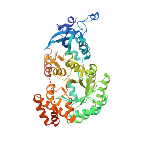
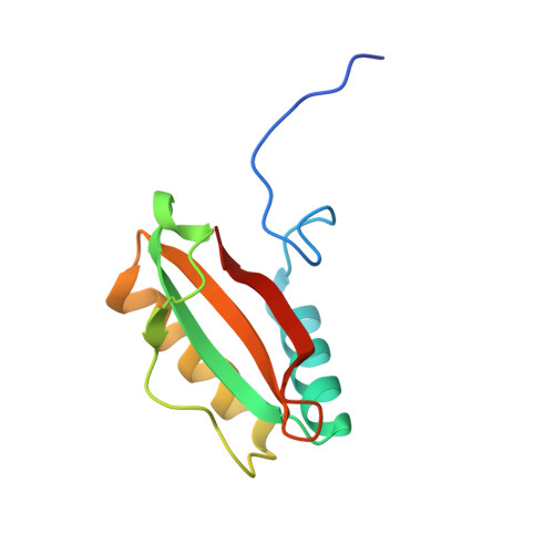
Multivalent interactions between CsoS2 and Rubisco mediate alpha-carboxysome formation.

Oltrogge, L.M.Chaijarasphong, T.Chen, A.W.Bolin, E.R.Marqusee, S.Savage, D.F.

(2020) Nat Struct Mol Biol 27: 281-287

  • DOI: https://doi.org/10.1038/s41594-020-0387-7
  • Primary Citation Related Structures: 
    6UEW

  • PubMed Abstract: 

    Carboxysomes are bacterial microcompartments that function as the centerpiece of the bacterial CO 2 -concentrating mechanism by facilitating high CO 2 concentrations near the carboxylase Rubisco. The carboxysome self-assembles from thousands of individual proteins into icosahedral-like particles with a dense enzyme cargo encapsulated within a proteinaceous shell. In the case of the α-carboxysome, there is little molecular insight into protein-protein interactions that drive the assembly process. Here, studies on the α-carboxysome from Halothiobacillus neapolitanus demonstrate that Rubisco interacts with the N terminus of CsoS2, a multivalent, intrinsically disordered protein. X-ray structural analysis of the CsoS2 interaction motif bound to Rubisco reveals a series of conserved electrostatic interactions that are only made with properly assembled hexadecameric Rubisco. Although biophysical measurements indicate that this single interaction is weak, its implicit multivalency induces high-affinity binding through avidity. Taken together, our results indicate that CsoS2 acts as an interaction hub to condense Rubisco and enable efficient α-carboxysome formation.


  • Organizational Affiliation
    • Department of Molecular and Cell Biology, University of California Berkeley, Berkeley, CA, USA.

Macromolecule Content 

  • Total Structure Weight: 276.63 kDa 
  • Atom Count: 18,304 
  • Modeled Residue Count: 2,228 
  • Deposited Residue Count: 2,464 
  • Unique protein chains: 2

Macromolecules

Find similar proteins by:|  3D Structure
Entity ID: 1
MoleculeChains  Sequence LengthOrganismDetailsImage
Ribulose bisphosphate carboxylase large chain, CsoS2 N-peptide fusion
A, C, E, G
506Halothiobacillus neapolitanus c2Mutation(s): 0 
Gene Names: cbbLHneap_0922
EC: 4.1.1.39
UniProt
Find proteins for O85040 (Halothiobacillus neapolitanus (strain ATCC 23641 / DSM 15147 / CIP 104769 / NCIMB 8539 / c2))
Explore O85040 
Go to UniProtKB:  O85040
Entity Groups
Sequence Clusters30% Identity50% Identity70% Identity90% Identity95% Identity100% Identity
UniProt GroupO85040
Sequence Annotations
Expand
Reference Sequence
Find similar proteins by:|  3D Structure
Entity ID: 2
MoleculeChains  Sequence LengthOrganismDetailsImage
Ribulose bisphosphate carboxylase small chain
B, D, F, H
110Halothiobacillus neapolitanus c2Mutation(s): 0 
Gene Names: cbbSrbcSHneap_0921
EC: 4.1.1.39
UniProt
Find proteins for P45686 (Halothiobacillus neapolitanus (strain ATCC 23641 / DSM 15147 / CIP 104769 / NCIMB 8539 / c2))
Explore P45686 
Go to UniProtKB:  P45686
Entity Groups
Sequence Clusters30% Identity50% Identity70% Identity90% Identity95% Identity100% Identity
UniProt GroupP45686
Sequence Annotations
Expand
Reference Sequence

Experimental Data & Validation

Experimental Data

  • Method: X-RAY DIFFRACTION
  • Resolution: 2.40 Å
  • R-Value Free:  0.259 (Depositor), 0.261 (DCC) 
  • R-Value Work:  0.188 (Depositor), 0.192 (DCC) 
  • R-Value Observed: 0.189 (Depositor) 
Space Group: C 1 2 1
Unit Cell:
Length ( Å )Angle ( ˚ )
a = 171.831α = 90
b = 153.945β = 124.703
c = 108.063γ = 90
Software Package:
Software NamePurpose
PHENIXrefinement
Blu-Icedata collection
XDSdata reduction
Aimlessdata scaling
Cootmodel building
PHENIXphasing

Structure Validation

View Full Validation Report


Entry History 

& Funding Information

Deposition Data


Funding OrganizationLocationGrant Number
National Institutes of Health/National Institute of General Medical Sciences (NIH/NIGMS)United StatesR01GM129241

Revision History  (Full details and data files)

  • Version 1.0: 2019-10-30
    Type: Initial release
  • Version 1.1: 2020-01-01
    Changes: Author supporting evidence
  • Version 1.2: 2020-03-18
    Changes: Database references
  • Version 1.3: 2020-03-25
    Changes: Database references
  • Version 1.4: 2023-10-11
    Changes: Data collection, Database references, Refinement description