6UQK | pdb_00006uqk

Cryo-EM structure of type 3 IP3 receptor revealing presence of a self-binding peptide


Experimental Data Snapshot

  • Method: ELECTRON MICROSCOPY
  • Resolution: 3.77 Å
  • Aggregation State: PARTICLE 
  • Reconstruction Method: SINGLE PARTICLE 

wwPDB Validation 3D Report Full Report

Validation slider image for 6UQK

This is version 1.3 of the entry. See complete history

Literature

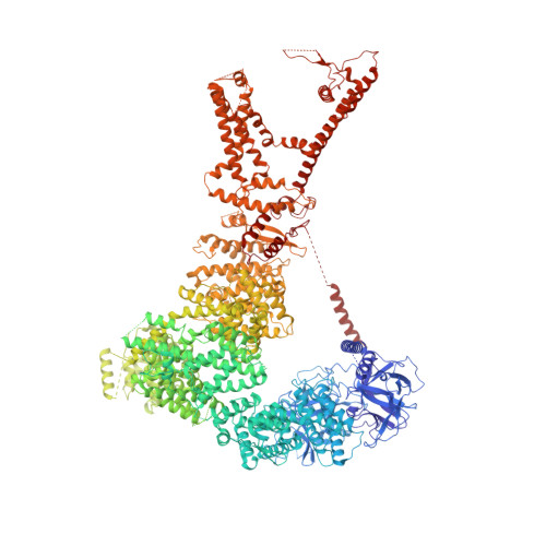
Cryo-EM structure of human type-3 inositol triphosphate receptor reveals the presence of a self-binding peptide that acts as an antagonist.

Azumaya, C.M.Linton, E.A.Risener, C.J.Nakagawa, T.Karakas, E.

(2020) J Biological Chem 295: 1743-1753

  • DOI: https://doi.org/10.1074/jbc.RA119.011570
  • Primary Citation Related Structures: 
    6UQK

  • PubMed Abstract: 

    Calcium-mediated signaling through inositol 1,4,5-triphosphate receptors (IP 3 Rs) is essential for the regulation of numerous physiological processes, including fertilization, muscle contraction, apoptosis, secretion, and synaptic plasticity. Deregulation of IP 3 Rs leads to pathological calcium signaling and is implicated in many common diseases, including cancer and neurodegenerative, autoimmune, and metabolic diseases. Revealing the mechanism of activation and inhibition of this ion channel will be critical to an improved understanding of the biological processes that are controlled by IP 3 Rs. Here, we report structural findings of the human type-3 IP 3 R (IP 3 R-3) obtained by cryo-EM (at an overall resolution of 3.8 Å), revealing an unanticipated regulatory mechanism where a loop distantly located in the primary sequence occupies the IP 3 -binding site and competitively inhibits IP 3 binding. We propose that this inhibitory mechanism must differ qualitatively among IP 3 R subtypes because of their diverse loop sequences, potentially serving as a key molecular determinant of subtype-specific calcium signaling in IP 3 Rs. In summary, our structural characterization of human IP 3 R-3 provides critical insights into the mechanistic function of IP 3 Rs and into subtype-specific regulation of these important calcium-regulatory channels.


  • Organizational Affiliation
    • Department of Molecular Physiology and Biophysics, Vanderbilt University, School of Medicine, Nashville, Tennessee 37232.

Macromolecule Content 

  • Total Structure Weight: 1,114.6 kDa 
  • Atom Count: 51,567 
  • Modeled Residue Count: 8,216 
  • Deposited Residue Count: 9,812 
  • Unique protein chains: 1

Macromolecules

Find similar proteins by:|  3D Structure
Entity ID: 1
MoleculeChains  Sequence LengthOrganismDetailsImage
inositol 1,4,5-triphosphate receptor, type 3
A, B, C, D
2,453Homo sapiensMutation(s): 0 
Gene Names: itpr3
Membrane Entity: Yes 
UniProt & NIH Common Fund Data Resources
Find proteins for Q14573 (Homo sapiens)
Explore Q14573 
Go to UniProtKB:  Q14573
PHAROS:  Q14573
GTEx:  ENSG00000096433 
Entity Groups
Sequence Clusters30% Identity50% Identity70% Identity90% Identity95% Identity100% Identity
UniProt GroupQ14573
Sequence Annotations
Expand
Reference Sequence

Experimental Data & Validation

Experimental Data

  • Method: ELECTRON MICROSCOPY
  • Resolution: 3.77 Å
  • Aggregation State: PARTICLE 
  • Reconstruction Method: SINGLE PARTICLE 
EM Software:
TaskSoftware PackageVersion
RECONSTRUCTIONcisTEM
MODEL REFINEMENTPHENIX

Structure Validation

View Full Validation Report


Entry History 

& Funding Information

Deposition Data


Funding OrganizationLocationGrant Number
National Institutes of Health/National Institute of Diabetes and Digestive and Kidney Disease (NIH/NIDDK)United StatesDK020593

Revision History  (Full details and data files)

  • Version 1.0: 2020-01-15
    Type: Initial release
  • Version 1.1: 2020-01-22
    Changes: Database references
  • Version 1.2: 2020-02-19
    Changes: Database references
  • Version 1.3: 2024-03-20
    Changes: Data collection, Database references