


Crystal Structure of Human Cathepsin-G Inhibited by S. aureus EapH1
Herdendorf, T.J., Rooijakkers, S.H.M., Geisbrecht, B.V.(2020) J Biological Chem
Experimental Data Snapshot
Starting Model: experimental
View more details
wwPDB Validation 3D Report Full Report

(2020) J Biological Chem
Entity ID: 1 | |||||
|---|---|---|---|---|---|
| Molecule | Chains | Sequence Length | Organism | Details | Image |
| Cathepsin G | 224 | Homo sapiens | Mutation(s): 0 EC: 3.4.21.20 | 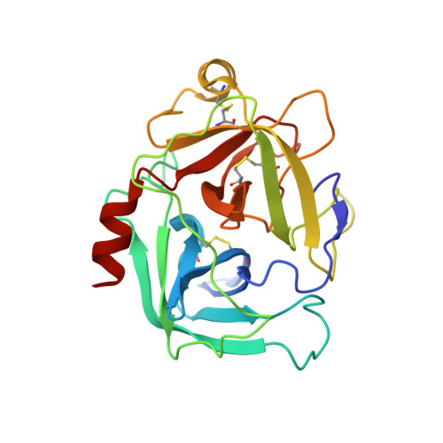 | |
UniProt & NIH Common Fund Data Resources | |||||
PHAROS: P08311 GTEx: ENSG00000100448 | |||||
Entity Groups | |||||
| Sequence Clusters | 30% Identity50% Identity70% Identity90% Identity95% Identity100% Identity | ||||
| UniProt Group | P08311 | ||||
Sequence AnnotationsExpand | |||||
Reference Sequence | |||||
Entity ID: 2 | |||||
|---|---|---|---|---|---|
| Molecule | Chains | Sequence Length | Organism | Details | Image |
| MAP domain-containing protein | 99 | Staphylococcus aureus subsp. aureus Mu50 | Mutation(s): 0 Gene Names: SAV2205 | 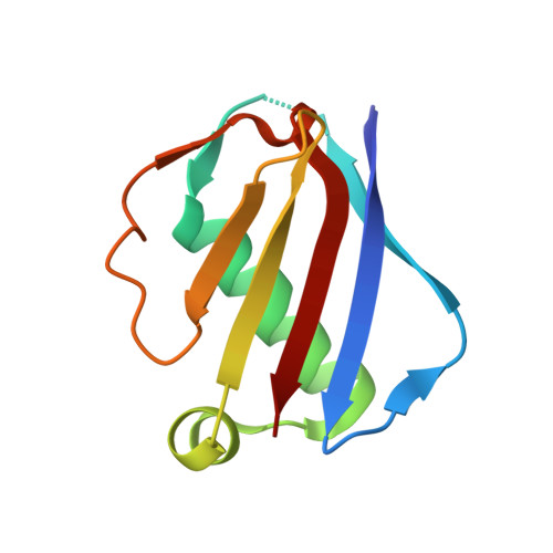 | |
UniProt | |||||
Find proteins for A0A0H3K0M1 (Staphylococcus aureus (strain Mu50 / ATCC 700699)) Explore A0A0H3K0M1 Go to UniProtKB: A0A0H3K0M1 | |||||
Entity Groups | |||||
| Sequence Clusters | 30% Identity50% Identity70% Identity90% Identity95% Identity100% Identity | ||||
| UniProt Group | A0A0H3K0M1 | ||||
Sequence AnnotationsExpand | |||||
Reference Sequence | |||||
| Ligands 1 Unique | |||||
|---|---|---|---|---|---|
| ID | Chains | Name / Formula / InChI Key | 2D Diagram | 3D Interactions | |
| SO4 Download:Ideal Coordinates CCD File | I [auth A] J [auth A] K [auth A] L [auth A] M [auth C] | SULFATE ION O4 S QAOWNCQODCNURD-UHFFFAOYSA-L |  | ||
| Length ( Å ) | Angle ( ˚ ) |
|---|---|
| a = 59.229 | α = 113.58 |
| b = 72.902 | β = 91.17 |
| c = 88.906 | γ = 106.24 |
| Software Name | Purpose |
|---|---|
| PHENIX | refinement |
| HKL-2000 | data reduction |
| HKL-2000 | data scaling |
| PHASER | phasing |
| PDB_EXTRACT | data extraction |
| Funding Organization | Location | Grant Number |
|---|---|---|
| National Institutes of Health/National Institute of General Medical Sciences (NIH/NIGMS) | United States | GM121511 |


