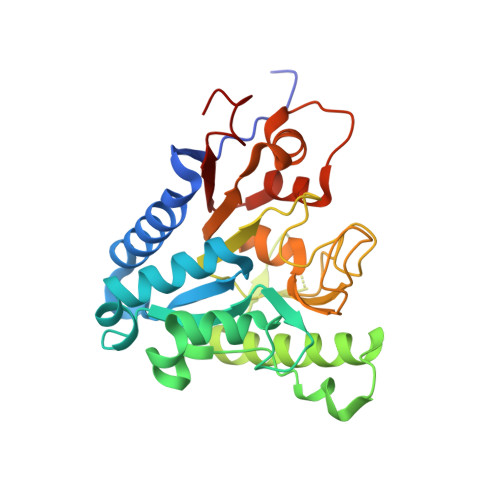
6VYE | pdb_00006vye

6-phosphogluconolactonase from Trypanosoma cruzi


Experimental Data Snapshot

  • Method: X-RAY DIFFRACTION
  • Resolution: 1.95 Å
  • R-Value Free: 
    0.270 (Depositor), 0.281 (DCC) 
  • R-Value Work: 
    0.231 (Depositor), 0.244 (DCC) 

Starting Model: experimental
View more details

wwPDB Validation 3D Report Full Report

Validation slider image for 6VYE

This is version 1.1 of the entry. See complete history

Literature

Macromolecule Content 

  • Total Structure Weight: 59.97 kDa 
  • Atom Count: 4,317 
  • Modeled Residue Count: 530 
  • Deposited Residue Count: 542 
  • Unique protein chains: 1

Macromolecules

Find similar proteins by:|  3D Structure
Entity ID: 1
MoleculeChains  Sequence LengthOrganismDetailsImage
6-phosphogluconolactonase
A, B
271Trypanosoma cruzi strain CL BrenerMutation(s): 0 
Gene Names: Tc00.1047053503945.40
EC: 3.1.1.31
UniProt
Find proteins for Q4CQA8 (Trypanosoma cruzi (strain CL Brener))
Explore Q4CQA8 
Go to UniProtKB:  Q4CQA8
Entity Groups
Sequence Clusters30% Identity50% Identity70% Identity90% Identity95% Identity100% Identity
UniProt GroupQ4CQA8
Sequence Annotations
Expand
Reference Sequence

Experimental Data & Validation

Experimental Data

  • Method: X-RAY DIFFRACTION
  • Resolution: 1.95 Å
  • R-Value Free:  0.270 (Depositor), 0.281 (DCC) 
  • R-Value Work:  0.231 (Depositor), 0.244 (DCC) 
Space Group: P 1 2 1
Unit Cell:
Length ( Å )Angle ( ˚ )
a = 71.77α = 90
b = 63.39β = 94.91
c = 79.32γ = 90
Software Package:
Software NamePurpose
MOSFLMdata reduction
Aimlessdata scaling
REFMACrefinement
PDB_EXTRACTdata extraction
MoRDaphasing

Structure Validation

View Full Validation Report


Entry History 

Deposition Data

Revision History  (Full details and data files)

  • Version 1.0: 2020-03-18
    Type: Initial release
  • Version 1.1: 2023-10-11
    Changes: Data collection, Database references, Refinement description