6WK1 | pdb_00006wk1

SETD3 in Complex with an Actin Peptide with His73 Replaced with Methionine


Experimental Data Snapshot

  • Method: X-RAY DIFFRACTION
  • Resolution: 1.89 Å
  • R-Value Free: 
    0.228 (Depositor), 0.230 (DCC) 
  • R-Value Work: 
    0.195 (Depositor), 0.196 (DCC) 
  • R-Value Observed: 
    0.195 (Depositor) 

Starting Model: experimental
View more details

wwPDB Validation 3D Report Full Report

Validation slider image for 6WK1

Ligand Structure Quality Assessment 


This is version 1.2 of the entry. See complete history

Literature

Characterization of SETD3 methyltransferase-mediated protein methionine methylation.

Dai, S.Holt, M.V.Horton, J.R.Woodcock, C.B.Patel, A.Zhang, X.Young, N.L.Wilkinson, A.W.Cheng, X.

(2020) J Biological Chem 295: 10901-10910

  • DOI: https://doi.org/10.1074/jbc.RA120.014072
  • Primary Citation Related Structures: 
    6WK1, 6WK2

  • PubMed Abstract: 

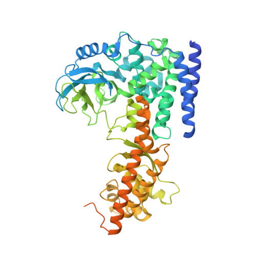
    Most characterized protein methylation events encompass arginine and lysine N -methylation, and only a few cases of protein methionine thiomethylation have been reported. Newly discovered oncohistone mutations include lysine-to-methionine substitutions at positions 27 and 36 of histone H3.3. In these instances, the methionine substitution localizes to the active-site pocket of the corresponding histone lysine methyltransferase, thereby inhibiting the respective transmethylation activity. SET domain-containing 3 (SETD3) is a protein ( i.e. actin) histidine methyltransferase. Here, we generated an actin variant in which the histidine target of SETD3 was substituted with methionine. As for previously characterized histone SET domain proteins, the methionine substitution substantially (76-fold) increased binding affinity for SETD3 and inhibited SETD3 activity on histidine. Unexpectedly, SETD3 was active on the substituted methionine, generating S -methylmethionine in the context of actin peptide. The ternary structure of SETD3 in complex with the methionine-containing actin peptide at 1.9 Å resolution revealed that the hydrophobic thioether side chain is packed by the aromatic rings of Tyr 312 and Trp 273 , as well as the hydrocarbon side chain of Ile 310 Our results suggest that placing methionine properly in the active site-within close proximity to and in line with the incoming methyl group of SAM-would allow some SET domain proteins to selectively methylate methionine in proteins.


  • Organizational Affiliation
    • Department of Epigenetics and Molecular Carcinogenesis, University of Texas M. D. Anderson Cancer Center, Houston, Texas, USA.

Macromolecule Content 

  • Total Structure Weight: 144.64 kDa 
  • Atom Count: 8,952 
  • Modeled Residue Count: 1,004 
  • Deposited Residue Count: 1,234 
  • Unique protein chains: 2

Macromolecules

Find similar proteins by:  Sequence   |   3D Structure  
Entity ID: 1
MoleculeChains  Sequence LengthOrganismDetailsImage
Actin, cytoplasmic 2A [auth Y],
C [auth Z]
23Homo sapiensMutation(s): 1 
EC: 3.6.4
UniProt & NIH Common Fund Data Resources
Find proteins for P63261 (Homo sapiens)
Explore P63261 
Go to UniProtKB:  P63261
PHAROS:  P63261
GTEx:  ENSG00000184009 
Entity Groups
Sequence Clusters30% Identity50% Identity70% Identity90% Identity95% Identity100% Identity
UniProt GroupP63261
Sequence Annotations
Expand
Reference Sequence
Find similar proteins by:|  3D Structure
Entity ID: 2
MoleculeChains  Sequence LengthOrganismDetailsImage
Actin-histidine N-methyltransferaseB [auth A],
D [auth B]
594Homo sapiensMutation(s): 0 
Gene Names: SETD3C14orf154
EC: 2.1.1.85
UniProt & NIH Common Fund Data Resources
Find proteins for Q86TU7 (Homo sapiens)
Explore Q86TU7 
Go to UniProtKB:  Q86TU7
PHAROS:  Q86TU7
GTEx:  ENSG00000183576 
Entity Groups
Sequence Clusters30% Identity50% Identity70% Identity90% Identity95% Identity100% Identity
UniProt GroupQ86TU7
Sequence Annotations
Expand
Reference Sequence

Small Molecules

Ligands 4 Unique
IDChains Name / Formula / InChI Key2D Diagram3D Interactions
SAH

Query on SAH


Download:Ideal Coordinates CCD File
G [auth A],
NA [auth B]
S-ADENOSYL-L-HOMOCYSTEINE
C14 H20 N6 O5 S
ZJUKTBDSGOFHSH-WFMPWKQPSA-N
GOL

Query on GOL


Download:Ideal Coordinates CCD File
E [auth Y]
H [auth A]
I [auth A]
J [auth A]
LA [auth A]
E [auth Y],
H [auth A],
I [auth A],
J [auth A],
LA [auth A],
MA [auth A]
GLYCEROL
C3 H8 O3
PEDCQBHIVMGVHV-UHFFFAOYSA-N
EDO

Query on EDO


Download:Ideal Coordinates CCD File
AA [auth A]
AB [auth B]
BA [auth A]
BB [auth B]
CA [auth A]
AA [auth A],
AB [auth B],
BA [auth A],
BB [auth B],
CA [auth A],
CB [auth B],
DB [auth B],
EA [auth A],
EB [auth B],
F [auth Y],
FA [auth A],
FB [auth B],
GA [auth A],
GB [auth B],
HA [auth A],
IA [auth A],
JA [auth A],
K [auth A],
KA [auth A],
L [auth A],
M [auth A],
N [auth A],
O [auth A],
OA [auth B],
P [auth A],
PA [auth B],
Q [auth A],
QA [auth B],
R [auth A],
RA [auth B],
S [auth A],
SA [auth B],
T [auth A],
TA [auth B],
U [auth A],
UA [auth B],
V [auth A],
VA [auth B],
W [auth A],
WA [auth B],
X [auth A],
XA [auth B],
Y [auth A],
YA [auth B],
Z [auth A],
ZA [auth B]
1,2-ETHANEDIOL
C2 H6 O2
LYCAIKOWRPUZTN-UHFFFAOYSA-N
ACT

Query on ACT


Download:Ideal Coordinates CCD File
DA [auth A]ACETATE ION
C2 H3 O2
QTBSBXVTEAMEQO-UHFFFAOYSA-M

Experimental Data & Validation

Experimental Data

  • Method: X-RAY DIFFRACTION
  • Resolution: 1.89 Å
  • R-Value Free:  0.228 (Depositor), 0.230 (DCC) 
  • R-Value Work:  0.195 (Depositor), 0.196 (DCC) 
  • R-Value Observed: 0.195 (Depositor) 
Space Group: P 1 21 1
Unit Cell:
Length ( Å )Angle ( ˚ )
a = 60.388α = 90
b = 175.531β = 92.359
c = 66.654γ = 90
Software Package:
Software NamePurpose
PHENIXrefinement
HKL-2000data reduction
HKL-2000data scaling
PHENIXphasing

Structure Validation

View Full Validation Report


Ligand Structure Quality Assessment 


Entry History 

& Funding Information

Deposition Data


Funding OrganizationLocationGrant Number
National Institutes of Health/National Institute of General Medical Sciences (NIH/NIGMS)United StatesGM049245-23

Revision History  (Full details and data files)

  • Version 1.0: 2020-06-17
    Type: Initial release
  • Version 1.1: 2020-08-19
    Changes: Database references
  • Version 1.2: 2023-10-18
    Changes: Data collection, Database references, Refinement description