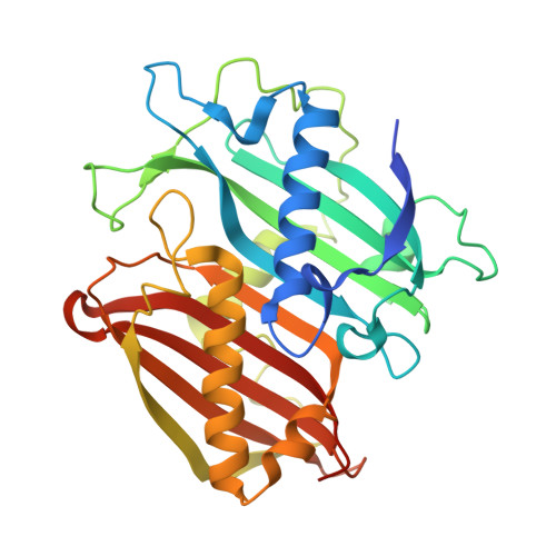
6X7M

LnmK in complex with 2-nitronate-propionyl-oxa(dethia)-CoA


Experimental Data Snapshot

  • Method: X-RAY DIFFRACTION
  • Resolution: 1.61 Å
  • R-Value Free: 0.227 
  • R-Value Work: 0.190 
  • R-Value Observed: 0.192 

Starting Model: experimental
View more details

wwPDB Validation   3D Report Full Report


Ligand Structure Quality Assessment 


This is version 1.3 of the entry. See complete history


Literature

Structures of LnmK, a Bifunctional Acyltransferase/Decarboxylase, with Substrate Analogues Reveal the Basis for Selectivity and Stereospecificity.

Stunkard, L.M.Kick, B.J.Lohman, J.R.

(2021) Biochemistry 60: 365-372

  • DOI: https://doi.org/10.1021/acs.biochem.0c00893
  • Primary Citation of Related Structures:  
    6X7L, 6X7M, 6X7N, 6X7O, 6X7P

  • PubMed Abstract: 

    LnmK stereospecifically accepts (2 R )-methylmalonyl-CoA, generating propionyl- S -acyl carrier protein to support polyketide biosynthesis. LnmK and its homologues are the only known enzymes that carry out a decarboxylation (DC) and acyl transfer (AT) reaction in the same active site as revealed by structure-function studies. Substrate-assisted catalysis powers LnmK, as decarboxylation of (2 R )-methylmalonyl-CoA generates an enolate capable of deprotonating active site Tyr62, and the Tyr62 phenolate subsequently attacks propionyl-CoA leading to a propionyl- O -LnmK acyl-enzyme intermediate. Due to the inherent reactivity of LnmK and methylmalonyl-CoA, a substrate-bound structure could not be obtained. To gain insight into substrate specificity, stereospecificity, and catalytic mechanism, we determined the structures of LnmK with bound substrate analogues that bear malonyl-thioester isosteres where the carboxylate is represented by a nitro or sulfonate group. The nitro-bearing malonyl-thioester isosteres bind in the nitronate form, with specific hydrogen bonds that allow modeling of the (2 R )-methylmalonyl-CoA substrate and rationalization of stereospecificity. The sulfonate isosteres bind in multiple conformations, suggesting the large active site of LnmK allows multiple binding modes. Considering the smaller malonyl group has more conformational freedom than the methylmalonyl group, we hypothesized the active site can entropically screen against catalysis with the smaller malonyl-CoA substrate. Indeed, our kinetic analysis reveals malonyl-CoA is accepted at 1% of the rate of methylmalonyl-CoA. This study represents another example of how our nitro- and sulfonate-bearing methylmalonyl-thioester isosteres are of use for elucidating enzyme-substrate binding interactions and revealing insights into catalytic mechanism. Synthesis of a larger panel of analogues presents an opportunity to study enzymes with complicated structure-function relationships such as acyl-CoA carboxylases, trans-carboxytransferases, malonyltransferases, and β-ketoacylsynthases.


  • Organizational Affiliation

    Department of Biochemistry, Purdue University, West Lafayette, Indiana 47907, United States.


Macromolecules
Find similar proteins by:  (by identity cutoff)  |  3D Structure
Entity ID: 1
MoleculeChains Sequence LengthOrganismDetailsImage
Bifunctional methylmalonyl-CoA:ACP acyltransferase/decarboxylase319Streptomyces atroolivaceusMutation(s): 1 
Gene Names: LnmK
UniProt
Find proteins for Q8GGP1 (Streptomyces atroolivaceus)
Explore Q8GGP1 
Go to UniProtKB:  Q8GGP1
Entity Groups  
Sequence Clusters30% Identity50% Identity70% Identity90% Identity95% Identity100% Identity
UniProt GroupQ8GGP1
Sequence Annotations
Expand
  • Reference Sequence
Small Molecules
Ligands 4 Unique
IDChains Name / Formula / InChI Key2D Diagram3D Interactions
KGA (Subject of Investigation/LOI)
Query on KGA

Download Ideal Coordinates CCD File 
B [auth A][1-[2-[3-[[(2~{R})-4-[[[(2~{R},3~{S},4~{R},5~{R})-5-(6-aminopurin-9-yl)-4-oxidanyl-3-phosphonooxy-oxolan-2-yl]methoxy-oxidanyl-phosphoryl]oxy-oxidanyl-phosphoryl]oxy-3,3-dimethyl-2-oxidanyl-butanoyl]amino]propanoylamino]ethoxy]-1-oxidanylidene-propan-2-ylidene]-bis(oxidanidyl)azanium
C24 H38 N8 O20 P3
JOSUZLPXQGIVES-ZSJPKINUSA-N
PGE
Query on PGE

Download Ideal Coordinates CCD File 
K [auth A]TRIETHYLENE GLYCOL
C6 H14 O4
ZIBGPFATKBEMQZ-UHFFFAOYSA-N
PEG
Query on PEG

Download Ideal Coordinates CCD File 
J [auth A]DI(HYDROXYETHYL)ETHER
C4 H10 O3
MTHSVFCYNBDYFN-UHFFFAOYSA-N
SO4
Query on SO4

Download Ideal Coordinates CCD File 
C [auth A]
D [auth A]
E [auth A]
F [auth A]
G [auth A]
C [auth A],
D [auth A],
E [auth A],
F [auth A],
G [auth A],
H [auth A],
I [auth A]
SULFATE ION
O4 S
QAOWNCQODCNURD-UHFFFAOYSA-L
Experimental Data & Validation

Experimental Data

  • Method: X-RAY DIFFRACTION
  • Resolution: 1.61 Å
  • R-Value Free: 0.227 
  • R-Value Work: 0.190 
  • R-Value Observed: 0.192 
  • Space Group: P 61 2 2
Unit Cell:
Length ( Å )Angle ( ˚ )
a = 61.55α = 90
b = 61.55β = 90
c = 308.805γ = 120
Software Package:
Software NamePurpose
SCALEPACKdata scaling
REFMACrefinement
PDB_EXTRACTdata extraction
REFMACphasing
DENZOdata reduction

Structure Validation

View Full Validation Report


Ligand Structure Quality Assessment 


Entry History 

Deposition Data

Revision History  (Full details and data files)

  • Version 1.0: 2020-06-10
    Type: Initial release
  • Version 1.1: 2021-02-03
    Changes: Database references
  • Version 1.2: 2021-02-17
    Changes: Database references
  • Version 1.3: 2023-10-18
    Changes: Data collection, Database references, Refinement description