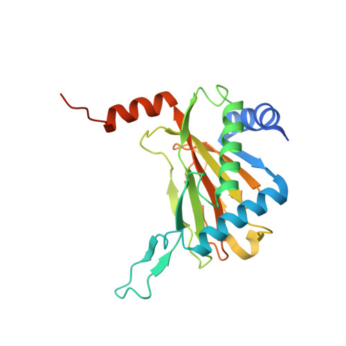
Use of cyclic peptides to induce crystallization: case study with prolyl hydroxylase domain 2.
Chowdhury, R., Abboud, M.I., McAllister, T.E., Banerji, B., Bhushan, B., Sorensen, J.L., Kawamura, A., Schofield, C.J.(2020) Sci Rep 10: 21964-21964
- PubMed: 33319810
- DOI: https://doi.org/10.1038/s41598-020-76307-8
- Primary Citation of Related Structures:
6YVW, 6YVX, 6YVZ, 6YW0, 6YW1, 6YW2, 6YW3, 6YW4 - PubMed Abstract:
Crystallization is the bottleneck in macromolecular crystallography; even when a protein crystallises, crystal packing often influences ligand-binding and protein-protein interaction interfaces, which are the key points of interest for functional and drug discovery studies. The human hypoxia-inducible factor prolyl hydroxylase 2 (PHD2) readily crystallises as a homotrimer, but with a sterically blocked active site. We explored strategies aimed at altering PHD2 crystal packing by protein modification and molecules that bind at its active site and elsewhere. Following the observation that, despite weak inhibition/binding in solution, succinamic acid derivatives readily enable PHD2 crystallization, we explored methods to induce crystallization without active site binding. Cyclic peptides obtained via mRNA display bind PHD2 tightly away from the active site. They efficiently enable PHD2 crystallization in different forms, both with/without substrates, apparently by promoting oligomerization involving binding to the C-terminal region. Although our work involves a specific case study, together with those of others, the results suggest that mRNA display-derived cyclic peptides may be useful in challenging protein crystallization cases.
Organizational Affiliation:
Chemistry Research Laboratory, Department of Chemistry, University of Oxford, Oxford, OX1 3TA, UK.


