6ZK8 | pdb_00006zk8

Native crystal structure of anaerobic F420H2-Oxidase from Methanothermococcus thermolithotrophicus at 1.8A resolution


Experimental Data Snapshot

  • Method: X-RAY DIFFRACTION
  • Resolution: 1.83 Å
  • R-Value Free: 
    0.204 (Depositor), 0.200 (DCC) 
  • R-Value Work: 
    0.178 (Depositor), 0.180 (DCC) 
  • R-Value Observed: 
    0.179 (Depositor) 

Starting Model: experimental
View more details

wwPDB Validation   3D Report Full Report


Ligand Structure Quality Assessment 

Created with Raphaël 2.3.0Worse 01 BetterLigand structure goodness of fit to experimental dataBest fitted FMNClick on this verticalbar to view details

This is version 1.1 of the entry. See complete history


Literature

Krypton-derivatization highlights O 2 -channeling in a four-electron reducing oxidase.

Engilberge, S.Wagner, T.Carpentier, P.Girard, E.Shima, S.

(2020) Chem Commun (Camb) 56: 10863-10866

  • DOI: https://doi.org/10.1039/d0cc04557h
  • Primary Citation of Related Structures:  
    6ZK8, 6ZLF

  • PubMed Abstract: 

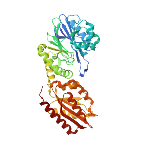
    F420H2-oxidase (FprA) catalyses the four-electron reduction of O2 to 2H2O using the reduced form of F420 as electron donor. The hydrophobic O2-channel detected by Kr-derivatization and the concerted movement of a gating loop could contribute to prevent unwanted side-reaction between the catalytic intermediates and solvents, therefore preventing reactive oxygen species formation.


  • Organizational Affiliation

    Paul Scherrer Institut, Forschungsstrasse 111, 5232 Villigen PSI, Switzerland.


Macromolecules
Find similar proteins by:  (by identity cutoff)  |  3D Structure
Entity ID: 1
MoleculeChains Sequence LengthOrganismDetailsImage
Coenzyme F420H2 oxidase (FprA)A,
B [auth O]
410Methanothermococcus thermolithotrophicusMutation(s): 0 
UniProt
Find proteins for A0A452CSW8 (Methanothermococcus thermolithotrophicus)
Explore A0A452CSW8 
Go to UniProtKB:  A0A452CSW8
Entity Groups  
Sequence Clusters30% Identity50% Identity70% Identity90% Identity95% Identity100% Identity
UniProt GroupA0A452CSW8
Sequence Annotations
Expand
  • Reference Sequence
Small Molecules
Ligands 5 Unique
IDChains Name / Formula / InChI Key2D Diagram3D Interactions
FMN
Query on FMN

Download Ideal Coordinates CCD File 
AA [auth O],
J [auth A]
FLAVIN MONONUCLEOTIDE
C17 H21 N4 O9 P
FVTCRASFADXXNN-SCRDCRAPSA-N
1PE
Query on 1PE

Download Ideal Coordinates CCD File 
F [auth A]PENTAETHYLENE GLYCOL
C10 H22 O6
JLFNLZLINWHATN-UHFFFAOYSA-N
PEG
Query on PEG

Download Ideal Coordinates CCD File 
C [auth A]
D [auth A]
E [auth A]
G [auth A]
H [auth A]
DI(HYDROXYETHYL)ETHER
C4 H10 O3
MTHSVFCYNBDYFN-UHFFFAOYSA-N
SO4
Query on SO4

Download Ideal Coordinates CCD File 
K [auth A]
L [auth A]
M [auth A]
N [auth A]
O [auth A]
SULFATE ION
O4 S
QAOWNCQODCNURD-UHFFFAOYSA-L
FE
Query on FE

Download Ideal Coordinates CCD File 
I [auth A],
P [auth A],
Y [auth O],
Z [auth O]
FE (III) ION
Fe
VTLYFUHAOXGGBS-UHFFFAOYSA-N
Experimental Data & Validation

Experimental Data

  • Method: X-RAY DIFFRACTION
  • Resolution: 1.83 Å
  • R-Value Free:  0.204 (Depositor), 0.200 (DCC) 
  • R-Value Work:  0.178 (Depositor), 0.180 (DCC) 
  • R-Value Observed: 0.179 (Depositor) 
Space Group: C 1 2 1
Unit Cell:
Length ( Å )Angle ( ˚ )
a = 100.871α = 90
b = 156.609β = 137.62
c = 76.065γ = 90
Software Package:
Software NamePurpose
PHENIXrefinement
XDSdata reduction
SCALAdata scaling
PHASERphasing
PDB_EXTRACTdata extraction

Structure Validation

View Full Validation Report


Ligand Structure Quality Assessment 

Created with Raphaël 2.3.0Worse 01 BetterLigand structure goodness of fit to experimental dataBest fitted FMNClick on this verticalbar to view details

Entry History & Funding Information

Deposition Data


Funding OrganizationLocationGrant Number
French National Research AgencyFranceLn23 ANR- 13-BS07-0007-01
Max Planck SocietyGermanyMax Planck Gessellschaft

Revision History  (Full details and data files)

  • Version 1.0: 2020-11-25
    Type: Initial release
  • Version 1.1: 2024-01-31
    Changes: Data collection, Database references, Refinement description