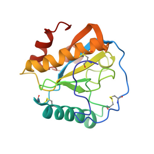
Crystal structure of the ternary complex of peptidoglycan recognition protein (PGRP-S) with Tartaric acid, Ribose and 2,6-DIAMINOPIMELIC ACID at 2.11 A resolution
Bairagya, H.R., Shokeen, A., Sharma, P., Singh, P.K., Sharma, S., Singh, T.P.To be published.