7A1X

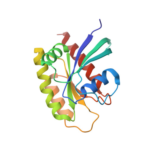
KRASG12C GDP form in complex with Cpd1


Experimental Data Snapshot

  • Method: X-RAY DIFFRACTION
  • Resolution: 1.32 Å
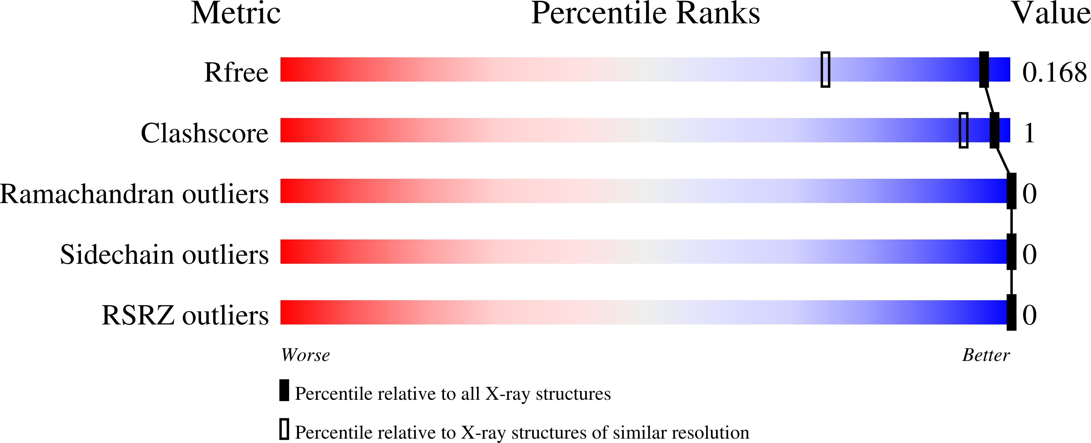
  • R-Value Free: 0.174 
  • R-Value Work: 0.148 

Starting Model: other
View more details

wwPDB Validation   3D Report Full Report


Ligand Structure Quality Assessment 


This is version 1.2 of the entry. See complete history


Literature

KRAS G12C fragment screening renders new binding pockets.

Mathieu, M.Steier, V.Fassy, F.Delorme, C.Papin, D.Genet, B.Duffieux, F.Bertrand, T.Delarbre, L.Le-Borgne, H.Parent, A.Didier, P.Marquette, J.P.Lowinski, M.Houtmann, J.Lamberton, A.Debussche, L.Alexey, R.

(2022) Small GTPases 13: 225-238

  • DOI: https://doi.org/10.1080/21541248.2021.1979360
  • Primary Citation of Related Structures:  
    7A1W, 7A1X, 7A1Y, 7A47

  • PubMed Abstract: 

    KRAS genes belong to the most frequently mutated family of oncogenes in cancer. The G12C mutation, found in a third of lung, half of colorectal and pancreatic cancer cases, is believed to be responsible for a substantial number of cancer deaths. For 30 years, KRAS has been the subject of extensive drug-targeting efforts aimed at targeting KRAS protein itself, but also its post-translational modifications, membrane localization, protein-protein interactions and downstream signalling pathways. So far, most KRAS targeting strategies have failed, and there are no KRAS-specific drugs available. However, clinical candidates targeting the KRAS G12C protein have recently been developed. MRTX849 and recently approved Sotorasib are covalent binders targeting the mutated cysteine 12, occupying Switch II pocket.Herein, we describe two fragment screening drug discovery campaigns that led to the identification of binding pockets on the KRAS G12C surface that have not previously been described. One screen focused on non-covalent binders to KRAS G12C, the other on covalent binders.


  • Organizational Affiliation

    Integrated Drug Discovery, Quai Jules Guesde, Vitry Sur Seine Cedex, France.


Macromolecules
Find similar proteins by:  (by identity cutoff)  |  3D Structure
Entity ID: 1
MoleculeChains Sequence LengthOrganismDetailsImage
GTPase KRas170Homo sapiensMutation(s): 1 
Gene Names: KRASKRAS2RASK2
EC: 3.6.5.2
UniProt & NIH Common Fund Data Resources
Find proteins for P01116 (Homo sapiens)
Go to UniProtKB:  P01116
PHAROS:  P01116
GTEx:  ENSG00000133703 
Sequence Annotations
Expand
  • Reference Sequence
Small Molecules
Ligands 3 Unique
IDChains Name / Formula / InChI Key2D Diagram3D Interactions
GDP
Query on GDP

Download Ideal Coordinates CCD File 
B [auth A]GUANOSINE-5'-DIPHOSPHATE
C10 H15 N5 O11 P2
QGWNDRXFNXRZMB-UUOKFMHZSA-N
QWB (Subject of Investigation/LOI)
Query on QWB

Download Ideal Coordinates CCD File 
D [auth A]3-(imidazol-1-ylmethyl)-1~{H}-indole
C12 H11 N3
FRADTKAUXQUOIJ-UHFFFAOYSA-N
MG
Query on MG

Download Ideal Coordinates CCD File 
C [auth A]MAGNESIUM ION
Mg
JLVVSXFLKOJNIY-UHFFFAOYSA-N
Experimental Data & Validation

Experimental Data

  • Method: X-RAY DIFFRACTION
  • Resolution: 1.32 Å
  • R-Value Free: 0.174 
  • R-Value Work: 0.148 
  • Space Group: P 21 21 21
Unit Cell:
Length ( Å )Angle ( ˚ )
a = 39.07α = 90
b = 40.69β = 90
c = 91.51γ = 90
Software Package:
Software NamePurpose
BUSTERrefinement
XDSdata reduction
Aimlessdata scaling
PHASERphasing

Structure Validation

View Full Validation Report


Ligand Structure Quality Assessment 


Entry History 

Deposition Data

Revision History  (Full details and data files)

  • Version 1.0: 2021-10-13
    Type: Initial release
  • Version 1.1: 2022-01-12
    Changes: Database references
  • Version 1.2: 2024-05-01
    Changes: Data collection, Refinement description