7A70 | pdb_00007a70

HEW lysozyme in complex with Ti(OH)4


Experimental Data Snapshot

  • Method: X-RAY DIFFRACTION
  • Resolution: 1.80 Å
  • R-Value Free: 
    0.239 (Depositor), 0.237 (DCC) 
  • R-Value Work: 
    0.194 (Depositor), 0.193 (DCC) 
  • R-Value Observed: 
    0.196 (Depositor) 

Starting Model: experimental
View more details

wwPDB Validation 3D Report Full Report

Validation slider image for 7A70

This is version 1.3 of the entry. See complete history

Literature

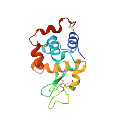
On the Mechanism of Bioinspired Formation of Inorganic Oxides: Structural Evidence of the Electrostatic Nature of the Interaction between a Mononuclear Inorganic Precursor and Lysozyme.

Gigli, L.Ravera, E.Calderone, V.Luchinat, C.

(2020) Biomolecules 11

  • DOI: https://doi.org/10.3390/biom11010043
  • Primary Citation Related Structures: 
    7A70

  • PubMed Abstract: 

    Nature has evolved several molecular machineries to promote the formation at physiological conditions of inorganic materials, which would otherwise be formed in extreme conditions. The molecular determinants of this process have been established over the last decade, identifying a strong role of electrostatics in the first steps of the precipitation. However, no conclusive, structure-based evidence has been provided so far. In this manuscript, we test the binding of lysozyme with silica and titania potential precursors. In contrast with the absence of structural information about the interaction with the silica precursor, we observe the interaction with a mononuclear titanium(IV) species, which is found to occur in a region rich of positive charges.


  • Organizational Affiliation
    • Magnetic Resonance Center (CERM), University of Florence, and Consorzio Interuniversitario Risonanze Magnetiche di Metalloproteine (CIRMMP), 50019 Sesto Fiorentino, Italy.

Macromolecule Content 

  • Total Structure Weight: 14.67 kDa 
  • Atom Count: 1,102 
  • Modeled Residue Count: 129 
  • Deposited Residue Count: 129 
  • Unique protein chains: 1

Macromolecules

Find similar proteins by:|  3D Structure
Entity ID: 1
MoleculeChains  Sequence LengthOrganismDetailsImage
Lysozyme129Gallus gallusMutation(s): 0 
EC: 3.2.1.17
UniProt
Find proteins for P00698 (Gallus gallus)
Explore P00698 
Go to UniProtKB:  P00698
Entity Groups
Sequence Clusters30% Identity50% Identity70% Identity90% Identity95% Identity100% Identity
UniProt GroupP00698
Sequence Annotations
Expand
Reference Sequence

Small Molecules

Ligands 4 Unique
IDChains Name / Formula / InChI Key2D Diagram3D Interactions
R3H
(Subject of Investigation/LOI)

Query on R3H


Download:Ideal Coordinates CCD File
G [auth A]tetrakis(oxidanyl)titanium
H4 O4 Ti
LLZRNZOLAXHGLL-UHFFFAOYSA-J
EDO

Query on EDO


Download:Ideal Coordinates CCD File
H [auth A]1,2-ETHANEDIOL
C2 H6 O2
LYCAIKOWRPUZTN-UHFFFAOYSA-N
CL

Query on CL


Download:Ideal Coordinates CCD File
C [auth A],
D [auth A],
E [auth A],
F [auth A]
CHLORIDE ION
Cl
VEXZGXHMUGYJMC-UHFFFAOYSA-M
NA

Query on NA


Download:Ideal Coordinates CCD File
B [auth A]SODIUM ION
Na
FKNQFGJONOIPTF-UHFFFAOYSA-N

Experimental Data & Validation

Experimental Data

  • Method: X-RAY DIFFRACTION
  • Resolution: 1.80 Å
  • R-Value Free:  0.239 (Depositor), 0.237 (DCC) 
  • R-Value Work:  0.194 (Depositor), 0.193 (DCC) 
  • R-Value Observed: 0.196 (Depositor) 
Space Group: P 43 21 2
Unit Cell:
Length ( Å )Angle ( ˚ )
a = 78.09α = 90
b = 78.09β = 90
c = 37.29γ = 90
Software Package:
Software NamePurpose
PHENIXrefinement
PDB_EXTRACTdata extraction
XDSdata reduction
XSCALEdata scaling
MOLREPphasing

Structure Validation

View Full Validation Report


Entry History 

Deposition Data

Revision History  (Full details and data files)

  • Version 1.0: 2021-01-13
    Type: Initial release
  • Version 1.1: 2021-01-20
    Changes: Database references
  • Version 1.2: 2024-01-31
    Changes: Data collection, Database references, Refinement description
  • Version 1.3: 2024-10-16
    Changes: Structure summary