7AZ9

Perdeuterated E65Q-TIM complexed with PHOSPHOGLYCOLOHYDROXAMATE


Experimental Data Snapshot

  • Method: X-RAY DIFFRACTION
  • Resolution: 1.10 Å
  • R-Value Free: 0.158 
  • R-Value Work: 0.144 
  • R-Value Observed: 0.144 

  • Method: NEUTRON DIFFRACTION
  • Resolution: 1.80 Å
  • R-Value Free: 0.259 
  • R-Value Work: 0.220 
  • R-Value Observed: 0.222 

wwPDB Validation   3D Report Full Report


This is version 1.0 of the entry. See complete history


Literature

Neutron structures of Leishmania mexicana triosephosphate isomerase in complex with reaction-intermediate mimics shed light on the proton-shuttling steps.

Kelpsas, V.Caldararu, O.Blakeley, M.P.Coquelle, N.Wierenga, R.K.Ryde, U.von Wachenfeldt, C.Oksanen, E.

(2021) IUCrJ 8: 633-643

  • DOI: https://doi.org/10.1107/S2052252521004619
  • Primary Citation of Related Structures:  
    7ABX, 7AZ3, 7AZ4, 7AZ9, 7AZA

  • PubMed Abstract: 

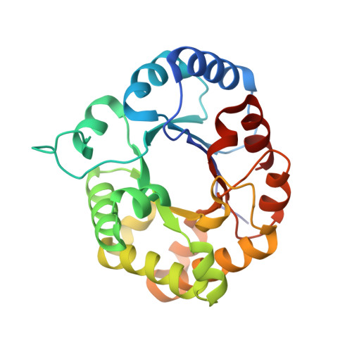
    Triosephosphate isomerase (TIM) is a key enzyme in glycolysis that catalyses the interconversion of glyceraldehyde 3-phosphate and dihydroxy-acetone phosphate. This simple reaction involves the shuttling of protons mediated by protolysable side chains. The catalytic power of TIM is thought to stem from its ability to facilitate the deprotonation of a carbon next to a carbonyl group to generate an enediolate intermediate. The enediolate intermediate is believed to be mimicked by the inhibitor 2-phosphoglycolate (PGA) and the subsequent enediol intermediate by phosphoglycolohydroxamate (PGH). Here, neutron structures of Leishmania mexicana TIM have been determined with both inhibitors, and joint neutron/X-ray refinement followed by quantum refinement has been performed. The structures show that in the PGA complex the postulated general base Glu167 is protonated, while in the PGH complex it remains deprotonated. The deuteron is clearly localized on Glu167 in the PGA-TIM structure, suggesting an asymmetric hydrogen bond instead of a low-barrier hydrogen bond. The full picture of the active-site protonation states allowed an investigation of the reaction mechanism using density-functional theory calculations.


  • Organizational Affiliation

    Department of Biology, Lund University, Sölvegatan 35, 223 62 Lund, Sweden.


Macromolecules
Find similar proteins by:  (by identity cutoff)  |  3D Structure
Entity ID: 1
MoleculeChains Sequence LengthOrganismDetailsImage
Triosephosphate isomerase251Leishmania mexicanaMutation(s): 1 
EC: 5.3.1.1
UniProt
Find proteins for P48499 (Leishmania mexicana)
Explore P48499 
Go to UniProtKB:  P48499
Entity Groups  
Sequence Clusters30% Identity50% Identity70% Identity90% Identity95% Identity100% Identity
UniProt GroupP48499
Sequence Annotations
Expand
  • Reference Sequence
Small Molecules
Ligands 1 Unique
IDChains Name / Formula / InChI Key2D Diagram3D Interactions
PGH (Subject of Investigation/LOI)
Query on PGH

Download Ideal Coordinates CCD File 
B [auth A]PHOSPHOGLYCOLOHYDROXAMIC ACID
C2 H6 N O6 P
BAXHHWZKQZIJID-UHFFFAOYSA-N
Experimental Data & Validation

Experimental Data

  • Method: X-RAY DIFFRACTION
  • Resolution: 1.10 Å
  • R-Value Free: 0.158 
  • R-Value Work: 0.144 
  • R-Value Observed: 0.144 
  • Space Group: C 1 2 1
  • Method: NEUTRON DIFFRACTION
  • Resolution: 1.80 Å
  • R-Value Free: 0.259 
  • R-Value Work: 0.220 
  • R-Value Observed: 0.222 
  • Space Group: C 1 2 1
Unit Cell:
Length ( Å )Angle ( ˚ )
a = 98.323α = 90
b = 52.678β = 117.86
c = 58.665γ = 90
Software Package:
Software NamePurpose
PHENIXrefinement
XDSdata reduction
LAUEGENdata reduction
XSCALEdata scaling
SCALAdata scaling
PHASERphasing

Structure Validation

View Full Validation Report


Entry History & Funding Information

Deposition Data


Funding OrganizationLocationGrant Number
Other privateSweden--
Other governmentSweden--

Revision History  (Full details and data files)

  • Version 1.0: 2021-07-28
    Type: Initial release