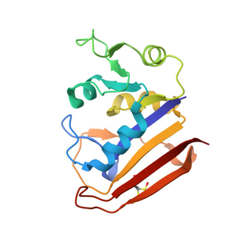
7FQE

DHFR:NADP+:FOL complex (crystal 2, pass 3, 295 K)


Experimental Data Snapshot

  • Method: X-RAY DIFFRACTION
  • Resolution: 1.18 Å
  • R-Value Free: 0.164 
  • R-Value Work: 0.129 
  • R-Value Observed: 0.131 

wwPDB Validation   3D Report Full Report


Ligand Structure Quality Assessment 


This is version 1.2 of the entry. See complete history


Literature

Perturbative diffraction methods resolve a conformational switch that facilitates a two-step enzymatic mechanism.

Greisman, J.B.Dalton, K.M.Brookner, D.E.Klureza, M.A.Sheehan, C.J.Kim, I.S.Henning, R.W.Russi, S.Hekstra, D.R.

(2024) Proc Natl Acad Sci U S A 121: e2313192121-e2313192121

  • DOI: https://doi.org/10.1073/pnas.2313192121
  • Primary Citation of Related Structures:  
    5SSS, 5SST, 5SSU, 5SSV, 5SSW, 7FPL, 7FPM, 7FPN, 7FPO, 7FPP, 7FPQ, 7FPR, 7FPS, 7FPT, 7FPU, 7FPV, 7FPW, 7FPX, 7FPY, 7FPZ, 7FQ0, 7FQ1, 7FQ2, 7FQ3, 7FQ4, 7FQ5, 7FQ6, 7FQ7, 7FQ8, 7FQ9, 7FQA, 7FQB, 7FQC, 7FQD, 7FQE, 7FQF, 7FQG, 8DAI, 8G4Z, 8G50

  • PubMed Abstract: 

    Enzymes catalyze biochemical reactions through precise positioning of substrates, cofactors, and amino acids to modulate the transition-state free energy. However, the role of conformational dynamics remains poorly understood due to poor experimental access. This shortcoming is evident with Escherichia coli dihydrofolate reductase (DHFR), a model system for the role of protein dynamics in catalysis, for which it is unknown how the enzyme regulates the different active site environments required to facilitate proton and hydride transfer. Here, we describe ligand-, temperature-, and electric-field-based perturbations during X-ray diffraction experiments to map the conformational dynamics of the Michaelis complex of DHFR. We resolve coupled global and local motions and find that these motions are engaged by the protonated substrate to promote efficient catalysis. This result suggests a fundamental design principle for multistep enzymes in which pre-existing dynamics enable intermediates to drive rapid electrostatic reorganization to facilitate subsequent chemical steps.


  • Organizational Affiliation

    Department of Molecular & Cellular Biology, Harvard University, Cambridge, MA 02138.


Macromolecules
Find similar proteins by:  (by identity cutoff)  |  3D Structure
Entity ID: 1
MoleculeChains Sequence LengthOrganismDetailsImage
Dihydrofolate reductase159Escherichia coli K-12Mutation(s): 0 
Gene Names: folAtmrAb0048JW0047
EC: 1.5.1.3
UniProt
Find proteins for P0ABQ4 (Escherichia coli (strain K12))
Explore P0ABQ4 
Go to UniProtKB:  P0ABQ4
Entity Groups  
Sequence Clusters30% Identity50% Identity70% Identity90% Identity95% Identity100% Identity
UniProt GroupP0ABQ4
Sequence Annotations
Expand
  • Reference Sequence
Small Molecules
Modified Residues  1 Unique
IDChains TypeFormula2D DiagramParent
CSD
Query on CSD
A
L-PEPTIDE LINKINGC3 H7 N O4 SCYS
Experimental Data & Validation

Experimental Data

  • Method: X-RAY DIFFRACTION
  • Resolution: 1.18 Å
  • R-Value Free: 0.164 
  • R-Value Work: 0.129 
  • R-Value Observed: 0.131 
  • Space Group: P 21 21 21
Unit Cell:
Length ( Å )Angle ( ˚ )
a = 34.248α = 90
b = 45.603β = 90
c = 99.094γ = 90
Software Package:
Software NamePurpose
DIALSdata reduction
DIALSdata scaling
PHENIXrefinement
PHENIXphasing

Structure Validation

View Full Validation Report


Ligand Structure Quality Assessment 


Entry History & Funding Information

Deposition Data


Funding OrganizationLocationGrant Number
Searle Scholars ProgramUnited StatesSSP-2018-3240
George W. Merck Fund in the New York Community TrustUnited States338034
National Institutes of Health/Office of the DirectorUnited StatesDP2-GM141000
National Science Foundation (NSF, United States)United StatesDGE1745303

Revision History  (Full details and data files)

  • Version 1.0: 2023-09-20
    Type: Initial release
  • Version 1.1: 2024-07-24
    Changes: Database references
  • Version 1.2: 2024-11-20
    Changes: Structure summary