7JFM | pdb_00007jfm

Crystal structure of mouse phosphorylated IRF-3 bound to CBP


Experimental Data Snapshot

  • Method: X-RAY DIFFRACTION
  • Resolution: 2.23 Å
  • R-Value Free: 
    0.252 (Depositor), 0.254 (DCC) 
  • R-Value Work: 
    0.231 (Depositor), 0.235 (DCC) 
  • R-Value Observed: 
    0.232 (Depositor) 

Starting Model: experimental
View more details

wwPDB Validation 3D Report Full Report

Validation slider image for 7JFM

This is version 1.3 of the entry. See complete history

Literature

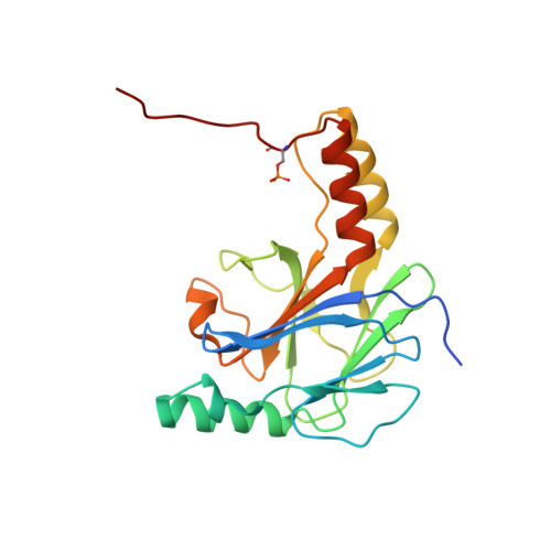
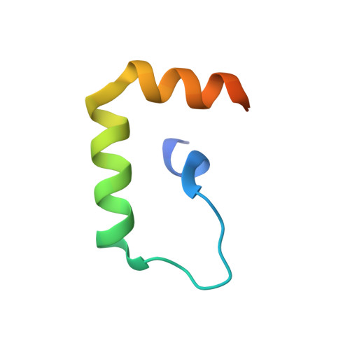
The Structural Basis of IRF-3 Activation upon Phosphorylation.

Jing, T.Zhao, B.Xu, P.Gao, X.Chi, L.Han, H.Sankaran, B.Li, P.

(2020) J Immunol 205: 1886-1896

  • DOI: https://doi.org/10.4049/jimmunol.2000026
  • Primary Citation Related Structures: 
    7JFL, 7JFM

  • PubMed Abstract: 

    The innate immune system is the first line of defense against bacterial and viral infections. The recognition of pathogen-associated molecular patterns by the RIG-I-like receptors, TLRs, and cGAS leads to the induction of IFN-I by activating the transcription factor IRF-3. Although the mechanism of IRF-3 activation has been extensively studied, the structural basis of IRF-3 activation upon phosphorylation is not fully understood. In this study, we determined the crystal structures of phosphorylated human and mouse IRF-3 bound to CREB-binding protein (CBP), which reveal that phosphorylated IRF-3 forms a dimer via pSer 386 (pSer 379 in mouse IRF-3) and a downstream p L x IS motif. Size-exclusion chromatography and cell-based studies show that mutations of key residues interacting with pSer 386 severely impair IRF-3 activation and IFN-β induction. By contrast, phosphorylation of Ser 396 within the p L x IS motif of human IRF-3 only plays a moderate role in IRF-3 activation. The mouse IRF-3/CBP complex structure reveals that the mechanism of mouse IRF-3 activation is similar but distinct from human IRF-3. These structural and functional studies reveal the detailed mechanism of IRF-3 activation upon phosphorylation.


  • Organizational Affiliation
    • Department of Biochemistry and Biophysics, Texas A&M University, College Station, TX 77843.

Macromolecule Content 

  • Total Structure Weight: 57.85 kDa 
  • Atom Count: 3,715 
  • Modeled Residue Count: 468 
  • Deposited Residue Count: 514 
  • Unique protein chains: 2

Macromolecules

Find similar proteins by:|  3D Structure
Entity ID: 1
MoleculeChains  Sequence LengthOrganismDetailsImage
Interferon regulatory factor 3A,
C [auth B]
210Mus musculusMutation(s): 0 
Gene Names: Irf3
UniProt & NIH Common Fund Data Resources
Find proteins for P70671 (Mus musculus)
Explore P70671 
Go to UniProtKB:  P70671
IMPC:  MGI:1859179
Entity Groups
Sequence Clusters30% Identity50% Identity70% Identity90% Identity95% Identity100% Identity
UniProt GroupP70671
Sequence Annotations
Expand
Reference Sequence
Find similar proteins by:|  3D Structure
Entity ID: 2
MoleculeChains  Sequence LengthOrganismDetailsImage
CREB-binding proteinB [auth C],
D
47Homo sapiensMutation(s): 0 
Gene Names: CREBBPCBP
EC: 2.3.1.48 (PDB Primary Data), 2.3.1 (UniProt)
UniProt & NIH Common Fund Data Resources
Find proteins for Q92793 (Homo sapiens)
Explore Q92793 
Go to UniProtKB:  Q92793
PHAROS:  Q92793
GTEx:  ENSG00000005339 
Entity Groups
Sequence Clusters30% Identity50% Identity70% Identity90% Identity95% Identity100% Identity
UniProt GroupQ92793
Sequence Annotations
Expand
Reference Sequence

Small Molecules

Modified Residues  1 Unique
IDChains TypeFormula2D DiagramParent
SEP
Query on SEP
A,
C [auth B]
L-PEPTIDE LINKINGC3 H8 N O6 PSER

Experimental Data & Validation

Experimental Data

  • Method: X-RAY DIFFRACTION
  • Resolution: 2.23 Å
  • R-Value Free:  0.252 (Depositor), 0.254 (DCC) 
  • R-Value Work:  0.231 (Depositor), 0.235 (DCC) 
  • R-Value Observed: 0.232 (Depositor) 
Space Group: P 62
Unit Cell:
Length ( Å )Angle ( ˚ )
a = 118.801α = 90
b = 118.801β = 90
c = 72.17γ = 120
Software Package:
Software NamePurpose
HKL-2000data reduction
HKL-2000data scaling
PHASERphasing
PHENIXrefinement
PDB_EXTRACTdata extraction

Structure Validation

View Full Validation Report


Entry History 

& Funding Information

Deposition Data


Funding OrganizationLocationGrant Number
National Institutes of Health/National Institute Of Allergy and Infectious Diseases (NIH/NIAID)United StatesR01 AI145287

Revision History  (Full details and data files)

  • Version 1.0: 2020-09-09
    Type: Initial release
  • Version 1.1: 2020-10-07
    Changes: Database references
  • Version 1.2: 2023-10-18
    Changes: Data collection, Database references, Refinement description
  • Version 1.3: 2024-11-13
    Changes: Structure summary