7JRF

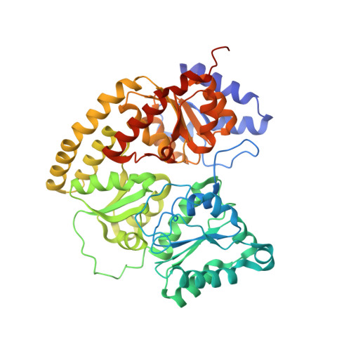
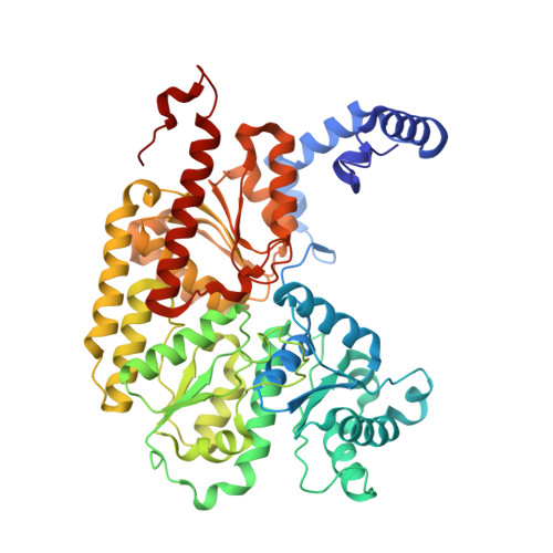
CO-CO-BOUND NITROGENASE MOFE-PROTEIN FROM A. VINELANDII


Experimental Data Snapshot

  • Method: X-RAY DIFFRACTION
  • Resolution: 1.33 Å
  • R-Value Free: 0.150 
  • R-Value Work: 0.116 
  • R-Value Observed: 0.118 

Starting Model: experimental
View more details

wwPDB Validation   3D Report Full Report


This is version 1.1 of the entry. See complete history


Literature

Structural Characterization of Two CO Molecules Bound to the Nitrogenase Active Site.

Buscagan, T.M.Perez, K.A.Maggiolo, A.O.Rees, D.C.Spatzal, T.

(2021) Angew Chem Int Ed Engl 60: 5704-5707

  • DOI: https://doi.org/10.1002/anie.202015751
  • Primary Citation of Related Structures:  
    7JRF

  • PubMed Abstract: 

    As an approach towards unraveling the nitrogenase mechanism, we have studied the binding of CO to the active-site FeMo-cofactor. CO is not only an inhibitor of nitrogenase, but it is also a substrate, undergoing reduction to hydrocarbons (Fischer-Tropsch-type chemistry). The C-C bond forming capabilities of nitrogenase suggest that multiple CO or CO-derived ligands bind to the active site. Herein, we report a crystal structure with two CO ligands coordinated to the FeMo-cofactor of the molybdenum nitrogenase at 1.33 Å resolution. In addition to the previously observed bridging CO ligand between Fe2 and Fe6 of the FeMo-cofactor, a new ligand binding mode is revealed through a second CO ligand coordinated terminally to Fe6. While the relevance of this state to nitrogenase-catalyzed reactions remains to be established, it highlights the privileged roles for Fe2 and Fe6 in ligand binding, with multiple coordination modes available depending on the ligand and reaction conditions.


  • Organizational Affiliation

    Division of Chemistry and Chemical Engineering, California Institute of Technology, 1200 E. California Blvd., Pasadena, CA, 91125, USA.


Macromolecules
Find similar proteins by:  (by identity cutoff)  |  3D Structure
Entity ID: 1
MoleculeChains Sequence LengthOrganismDetailsImage
Nitrogenase molybdenum-iron protein alpha chain
A, C
492Azotobacter vinelandiiMutation(s): 0 
EC: 1.18.6.1
UniProt
Find proteins for P07328 (Azotobacter vinelandii)
Go to UniProtKB:  P07328
Sequence Annotations
Expand
  • Reference Sequence
Find similar proteins by:  (by identity cutoff)  |  3D Structure
Entity ID: 2
MoleculeChains Sequence LengthOrganismDetailsImage
Nitrogenase molybdenum-iron protein beta chain
B, D
523Azotobacter vinelandiiMutation(s): 0 
EC: 1.18.6.1
UniProt
Find proteins for P07329 (Azotobacter vinelandii)
Go to UniProtKB:  P07329
Sequence Annotations
Expand
  • Reference Sequence
Small Molecules
Ligands 8 Unique
IDChains Name / Formula / InChI Key2D Diagram3D Interactions
ICE (Subject of Investigation/LOI)
Query on ICE

Download Ideal Coordinates CCD File 
F [auth A],
R [auth C]
iron-sulfur-molybdenum cluster with interstitial carbon
C Fe7 Mo S8
BJBPMDQXUSSDMI-UHFFFAOYSA-N
CLF
Query on CLF

Download Ideal Coordinates CCD File 
M [auth B],
X [auth D]
FE(8)-S(7) CLUSTER
Fe8 S7
JKVMXLBGZBULKV-UHFFFAOYSA-N
HCA
Query on HCA

Download Ideal Coordinates CCD File 
E [auth A],
Q [auth C]
3-HYDROXY-3-CARBOXY-ADIPIC ACID
C7 H10 O7
XKJVEVRQMLKSMO-SSDOTTSWSA-N
IMD
Query on IMD

Download Ideal Coordinates CCD File 
AA [auth D]
G [auth A]
H [auth A]
I [auth A]
O [auth B]
AA [auth D],
G [auth A],
H [auth A],
I [auth A],
O [auth B],
S [auth C],
Y [auth D],
Z [auth D]
IMIDAZOLE
C3 H5 N2
RAXXELZNTBOGNW-UHFFFAOYSA-O
CA
Query on CA

Download Ideal Coordinates CCD File 
N [auth B],
W [auth D]
CALCIUM ION
Ca
BHPQYMZQTOCNFJ-UHFFFAOYSA-N
H2S
Query on H2S

Download Ideal Coordinates CCD File 
J [auth A],
T [auth C]
HYDROSULFURIC ACID
H2 S
RWSOTUBLDIXVET-UHFFFAOYSA-N
CMO (Subject of Investigation/LOI)
Query on CMO

Download Ideal Coordinates CCD File 
K [auth A],
L [auth A],
U [auth C],
V [auth C]
CARBON MONOXIDE
C O
UGFAIRIUMAVXCW-UHFFFAOYSA-N
MG
Query on MG

Download Ideal Coordinates CCD File 
BA [auth D],
P [auth B]
MAGNESIUM ION
Mg
JLVVSXFLKOJNIY-UHFFFAOYSA-N
Experimental Data & Validation

Experimental Data

  • Method: X-RAY DIFFRACTION
  • Resolution: 1.33 Å
  • R-Value Free: 0.150 
  • R-Value Work: 0.116 
  • R-Value Observed: 0.118 
  • Space Group: P 1 21 1
Unit Cell:
Length ( Å )Angle ( ˚ )
a = 77.052α = 90
b = 129.974β = 109.11
c = 107.223γ = 90
Software Package:
Software NamePurpose
REFMACrefinement
PDB_EXTRACTdata extraction
XDSdata reduction
SCALAdata scaling
MOLREPphasing

Structure Validation

View Full Validation Report


Entry History 

Deposition Data

Revision History  (Full details and data files)

  • Version 1.0: 2021-03-24
    Type: Initial release
  • Version 1.1: 2023-10-18
    Changes: Data collection, Database references, Refinement description