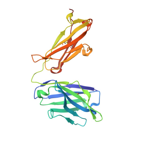
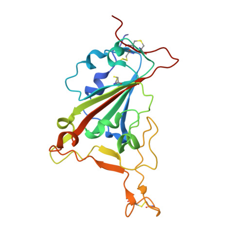
7JX3

Mapping neutralizing and immunodominant sites on the SARS-CoV-2 spike receptor-binding domain by structure-guided high-resolution serology


Experimental Data Snapshot

  • Method: X-RAY DIFFRACTION
  • Resolution: 2.65 Å
  • R-Value Free: 0.288 
  • R-Value Work: 0.258 
  • R-Value Observed: 0.259 

Starting Model: experimental
View more details

wwPDB Validation   3D Report Full Report


Ligand Structure Quality Assessment 


This is version 1.5 of the entry. See complete history


Literature

Mapping Neutralizing and Immunodominant Sites on the SARS-CoV-2 Spike Receptor-Binding Domain by Structure-Guided High-Resolution Serology.

Piccoli, L.Park, Y.J.Tortorici, M.A.Czudnochowski, N.Walls, A.C.Beltramello, M.Silacci-Fregni, C.Pinto, D.Rosen, L.E.Bowen, J.E.Acton, O.J.Jaconi, S.Guarino, B.Minola, A.Zatta, F.Sprugasci, N.Bassi, J.Peter, A.De Marco, A.Nix, J.C.Mele, F.Jovic, S.Rodriguez, B.F.Gupta, S.V.Jin, F.Piumatti, G.Lo Presti, G.Pellanda, A.F.Biggiogero, M.Tarkowski, M.Pizzuto, M.S.Cameroni, E.Havenar-Daughton, C.Smithey, M.Hong, D.Lepori, V.Albanese, E.Ceschi, A.Bernasconi, E.Elzi, L.Ferrari, P.Garzoni, C.Riva, A.Snell, G.Sallusto, F.Fink, K.Virgin, H.W.Lanzavecchia, A.Corti, D.Veesler, D.

(2020) Cell 183: 1024-1042.e21

  • DOI: https://doi.org/10.1016/j.cell.2020.09.037
  • Primary Citation of Related Structures:  
    7JV2, 7JV4, 7JV6, 7JVA, 7JVC, 7JW0, 7JX3, 7JXC, 7JXD, 7JXE

  • PubMed Abstract: 

    Analysis of the specificity and kinetics of neutralizing antibodies (nAbs) elicited by SARS-CoV-2 infection is crucial for understanding immune protection and identifying targets for vaccine design. In a cohort of 647 SARS-CoV-2-infected subjects, we found that both the magnitude of Ab responses to SARS-CoV-2 spike (S) and nucleoprotein and nAb titers correlate with clinical scores. The receptor-binding domain (RBD) is immunodominant and the target of 90% of the neutralizing activity present in SARS-CoV-2 immune sera. Whereas overall RBD-specific serum IgG titers waned with a half-life of 49 days, nAb titers and avidity increased over time for some individuals, consistent with affinity maturation. We structurally defined an RBD antigenic map and serologically quantified serum Abs specific for distinct RBD epitopes leading to the identification of two major receptor-binding motif antigenic sites. Our results explain the immunodominance of the receptor-binding motif and will guide the design of COVID-19 vaccines and therapeutics.


  • Organizational Affiliation

    Humabs BioMed SA, Vir Biotechnology, 6500 Bellinzona, Switzerland.


Macromolecules
Find similar proteins by:  (by identity cutoff)  |  3D Structure
Entity ID: 1
MoleculeChains Sequence LengthOrganismDetailsImage
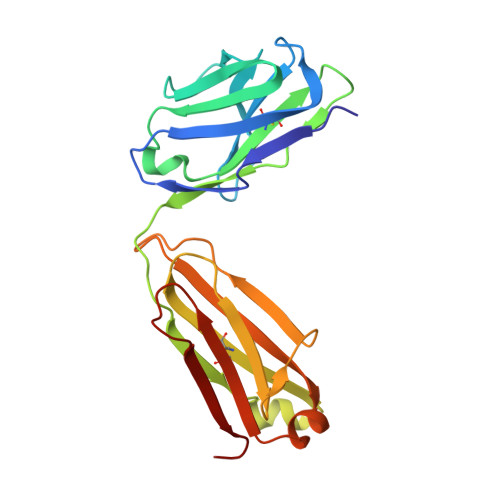
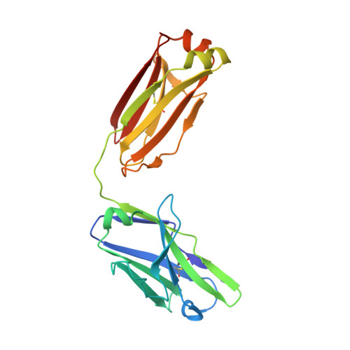
Light chain of Fab domain of monoclonal antibody S309A [auth B]214Homo sapiensMutation(s): 0 
Entity Groups  
Sequence Clusters30% Identity50% Identity70% Identity90% Identity95% Identity100% Identity
Sequence Annotations
Expand
  • Reference Sequence
Find similar proteins by:  (by identity cutoff)  |  3D Structure
Entity ID: 2
MoleculeChains Sequence LengthOrganismDetailsImage
Heavy chain of Fab domain of monoclonal antibody S309B [auth A]230Homo sapiensMutation(s): 0 
Entity Groups  
Sequence Clusters30% Identity50% Identity70% Identity90% Identity95% Identity100% Identity
Sequence Annotations
Expand
  • Reference Sequence
Find similar proteins by:  (by identity cutoff)  |  3D Structure
Entity ID: 3
MoleculeChains Sequence LengthOrganismDetailsImage
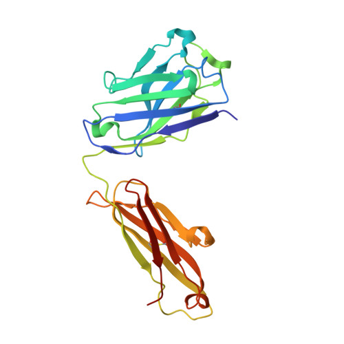
Light chain of Fab domain of monoclonal antibody S2H14C [auth D]216Homo sapiensMutation(s): 0 
Entity Groups  
Sequence Clusters30% Identity50% Identity70% Identity90% Identity95% Identity100% Identity
Sequence Annotations
Expand
  • Reference Sequence
Find similar proteins by:  (by identity cutoff)  |  3D Structure
Entity ID: 4
MoleculeChains Sequence LengthOrganismDetailsImage
Heavy chain of Fab domain of monoclonal antibody S2H14D [auth C]229Homo sapiensMutation(s): 0 
Entity Groups  
Sequence Clusters30% Identity50% Identity70% Identity90% Identity95% Identity100% Identity
Sequence Annotations
Expand
  • Reference Sequence
Find similar proteins by:  (by identity cutoff)  |  3D Structure
Entity ID: 5
MoleculeChains Sequence LengthOrganismDetailsImage
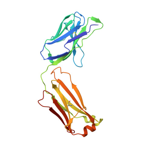
Light chain of Fab domain of monoclonal antibody S304E [auth L]215Homo sapiensMutation(s): 0 
Entity Groups  
Sequence Clusters30% Identity50% Identity70% Identity90% Identity95% Identity100% Identity
Sequence Annotations
Expand
  • Reference Sequence
Find similar proteins by:  (by identity cutoff)  |  3D Structure
Entity ID: 6
MoleculeChains Sequence LengthOrganismDetailsImage
Heavy chain of Fab domain of monoclonal antibody S304F [auth H]223Homo sapiensMutation(s): 0 
Entity Groups  
Sequence Clusters30% Identity50% Identity70% Identity90% Identity95% Identity100% Identity
Sequence Annotations
Expand
  • Reference Sequence
Find similar proteins by:  (by identity cutoff)  |  3D Structure
Entity ID: 7
MoleculeChains Sequence LengthOrganismDetailsImage
Spike protein S1G [auth R]204Severe acute respiratory syndrome coronavirus 2Mutation(s): 0 
Gene Names: S2
UniProt
Find proteins for P0DTC2 (Severe acute respiratory syndrome coronavirus 2)
Explore P0DTC2 
Go to UniProtKB:  P0DTC2
Entity Groups  
Sequence Clusters30% Identity50% Identity70% Identity90% Identity95% Identity100% Identity
UniProt GroupP0DTC2
Glycosylation
Glycosylation Sites: 1Go to GlyGen: P0DTC2-1
Sequence Annotations
Expand
  • Reference Sequence
Small Molecules
Ligands 1 Unique
IDChains Name / Formula / InChI Key2D Diagram3D Interactions
NAG
Query on NAG

Download Ideal Coordinates CCD File 
H [auth R]2-acetamido-2-deoxy-beta-D-glucopyranose
C8 H15 N O6
OVRNDRQMDRJTHS-FMDGEEDCSA-N
Experimental Data & Validation

Experimental Data

  • Method: X-RAY DIFFRACTION
  • Resolution: 2.65 Å
  • R-Value Free: 0.288 
  • R-Value Work: 0.258 
  • R-Value Observed: 0.259 
  • Space Group: C 1 2 1
Unit Cell:
Length ( Å )Angle ( ˚ )
a = 79.54α = 90
b = 127.78β = 96.66
c = 192.28γ = 90
Software Package:
Software NamePurpose
Aimlessdata scaling
PHASERphasing
REFMACrefinement
PDB_EXTRACTdata extraction
XDSdata reduction

Structure Validation

View Full Validation Report


Ligand Structure Quality Assessment 


Entry History & Funding Information

Deposition Data


Funding OrganizationLocationGrant Number
National Institutes of Health/National Institute of General Medical Sciences (NIH/NIGMS)United StatesGM120553
National Institutes of Health/National Institute Of Allergy and Infectious Diseases (NIH/NIAID)United StatesSeattle Structural Genomics Center for Infectious Diseases (SSGCID)

Revision History  (Full details and data files)

  • Version 1.0: 2020-10-14
    Type: Initial release
  • Version 1.1: 2020-11-25
    Changes: Database references
  • Version 1.2: 2021-01-27
    Changes: Structure summary
  • Version 1.3: 2023-10-18
    Changes: Author supporting evidence, Data collection, Database references, Refinement description, Structure summary
  • Version 1.4: 2024-08-07
    Changes: Other, Structure summary
  • Version 1.5: 2024-10-23
    Changes: Structure summary