A carboxylic acid isostere screen of the DHODH inhibitor Brequinar.
DeRatt, L.G., Christine Pietsch, E., Tanner, A., Shaffer, P., Jacoby, E., Wang, W., Kazmi, F., Zhang, X., Attar, R.M., Edwards, J.P., Kuduk, S.D.(2020) Bioorg Med Chem Lett 30: 127589-127589
- PubMed: 33007394 Search on PubMed
- DOI: https://doi.org/10.1016/j.bmcl.2020.127589
- Primary Citation Related Structures:
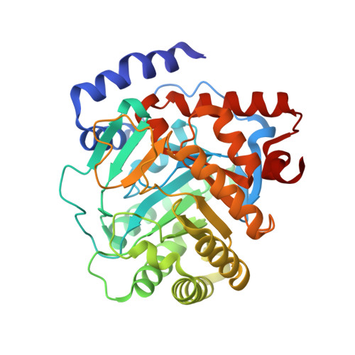
7K2U - PubMed Abstract:
Dihydroorotate dehydrogenase (DHODH) enzymatic activity impacts many aspects critical to cell proliferation and survival. Recently, DHODH has been identified as a target for acute myeloid differentiation therapy. In preclinical models of AML, the DHODH inhibitor Brequinar (BRQ) demonstrated potent anti-leukemic activity. Herein we describe a carboxylic acid isostere study of Brequinar which revealed a more potent non-carboxylic acid derivative with improved cellular potency and good pharmacokinetic properties.
- Discovery Chemistry, Janssen Pharmaceutical Research & Development, 1400 McKean Rd, Spring House, PA 19477, USA. Electronic address: lderatt@its.jnj.com.
Organizational Affiliation: