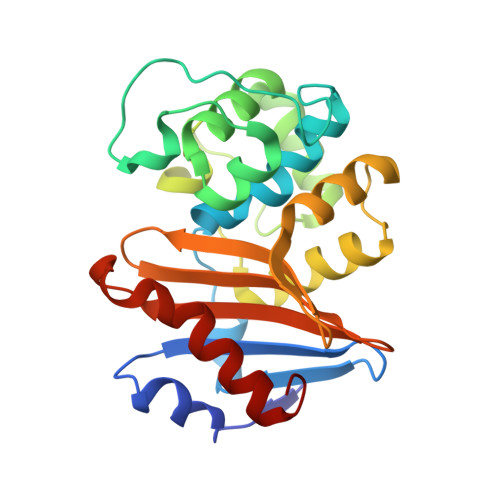
7KHZ | pdb_00007khz

Crystal structure of OXA-163 K73A in complex with imipenem


Experimental Data Snapshot

  • Method: X-RAY DIFFRACTION
  • Resolution: 2.04 Å
  • R-Value Free: 
    0.236 (Depositor), 0.240 (DCC) 
  • R-Value Work: 
    0.186 (Depositor), 0.190 (DCC) 
  • R-Value Observed: 
    0.188 (Depositor) 

Starting Model: experimental
View more details

wwPDB Validation   3D Report Full Report


Ligand Structure Quality Assessment 

Created with Raphaël 2.3.0Worse 01 BetterLigand structure goodness of fit to experimental dataBest fitted IM2Click on this verticalbar to view details

This is version 1.4 of the entry. See complete history


Literature

Mechanistic Basis of OXA-48-like beta-Lactamases' Hydrolysis of Carbapenems.

Stojanoski, V.Hu, L.Sankaran, B.Wang, F.Tao, P.Prasad, B.V.V.Palzkill, T.

(2021) ACS Infect Dis 7: 445-460

  • DOI: https://doi.org/10.1021/acsinfecdis.0c00798
  • Primary Citation of Related Structures:  
    7KH9, 7KHQ, 7KHY, 7KHZ

  • PubMed Abstract: 

    Carbapenem-hydrolyzing class D β-lactamases (CHDLs) are an important source of resistance to these last resort β-lactam antibiotics. OXA-48 is a member of a group of CHDLs named OXA-48-like enzymes. On the basis of sequence similarity, OXA-163 can be classified as an OXA-48-like enzyme, but it has altered substrate specificity. Compared to OXA-48, it shows impaired activity for carbapenems but displays an enhanced hydrolysis of oxyimino-cephalosporins. Here, we address the mechanistic and structural basis for carbapenem hydrolysis by OXA-48-like enzymes. Pre-steady-state kinetic analysis indicates that the rate-limiting step for OXA-48 and OXA-163 hydrolysis of carbapenems is deacylation and that the greatly reduced carbapenemase activity of OXA-163 compared to that of OXA-48 is due entirely to a slower deacylation reaction. Furthermore, our structural data indicate that the positioning of the β5-β6 loop is necessary for carbapenem hydrolysis by OXA-48. A major difference between the OXA-48 and OXA-163 complexes with carbapenems is that the 214-RIEP-217 deletion in OXA-163 creates a large opening in the active site that is absent in the OXA-48/carbapenem structures. We propose that the larger active site results in less constraint on the conformation of the 6α-hydroxyethyl group in the acyl-enzyme. The acyl-enzyme intermediate assumes multiple conformations, most of which are incompatible with rapid deacylation. Consistent with this hypothesis, molecular dynamics simulations indicate that the most stable complex is formed between OXA-48 and imipenem, which correlates with the OXA-48 hydrolysis of imipenem being the fastest observed. Furthermore, the OXA-163 complexes with imipenem and meropenem are the least stable and show significant conformational fluctuations, which correlates with the slow hydrolysis of these substrates.


  • Organizational Affiliation

    Department of Molecular Biophysics and Integrated Bioimaging, Berkeley Center for Structural Biology, Lawrence Berkeley National Laboratory, Berkeley, California 94720, United States.


Macromolecules
Find similar proteins by:  (by identity cutoff)  |  3D Structure
Entity ID: 1
MoleculeChains Sequence LengthOrganismDetailsImage
Beta-lactamase240Enterobacter cloacaeMutation(s): 1 
Gene Names: blaOXA-163
EC: 3.5.2.6
UniProt
Find proteins for F6KZJ2 (Enterobacter cloacae)
Explore F6KZJ2 
Go to UniProtKB:  F6KZJ2
Entity Groups  
Sequence Clusters30% Identity50% Identity70% Identity90% Identity95% Identity100% Identity
UniProt GroupF6KZJ2
Sequence Annotations
Expand
  • Reference Sequence
Experimental Data & Validation

Experimental Data

  • Method: X-RAY DIFFRACTION
  • Resolution: 2.04 Å
  • R-Value Free:  0.236 (Depositor), 0.240 (DCC) 
  • R-Value Work:  0.186 (Depositor), 0.190 (DCC) 
  • R-Value Observed: 0.188 (Depositor) 
Space Group: C 2 2 21
Unit Cell:
Length ( Å )Angle ( ˚ )
a = 44.42α = 90
b = 88.31β = 90
c = 125.35γ = 90
Software Package:
Software NamePurpose
PHENIXrefinement
MOSFLMdata reduction
SCALAdata scaling
MOLREPphasing

Structure Validation

View Full Validation Report


Ligand Structure Quality Assessment 

Created with Raphaël 2.3.0Worse 01 BetterLigand structure goodness of fit to experimental dataBest fitted IM2Click on this verticalbar to view details

Entry History & Funding Information

Deposition Data


Funding OrganizationLocationGrant Number
National Institutes of Health/National Institute Of Allergy and Infectious Diseases (NIH/NIAID)United States--

Revision History  (Full details and data files)

  • Version 1.0: 2021-02-10
    Type: Initial release
  • Version 1.1: 2021-02-24
    Changes: Database references
  • Version 1.2: 2021-06-30
    Changes: Structure summary
  • Version 1.3: 2023-10-18
    Changes: Data collection, Database references, Refinement description
  • Version 1.4: 2024-10-16
    Changes: Structure summary