Perturbing the Movement of Hydrogens to Delineate and Assign Events in the Reductive Activation and Turnover of Porcine Dihydropyrimidine Dehydrogenase.
Beaupre, B.A., Forouzesh, D.C., Butrin, A., Liu, D., Moran, G.R.(2021) Biochemistry 60: 1764-1775
- PubMed: 34032117 Search on PubMed
- DOI: https://doi.org/10.1021/acs.biochem.1c00243
- Primary Citation Related Structures:
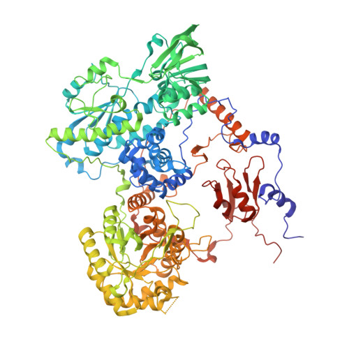
7M31, 7M32 - PubMed Abstract:
The native function of dihydropyrimidine dehydrogenase (DPD) is to reduce the 5,6-vinylic bond of pyrimidines uracil and thymine with electrons obtained from NADPH. NADPH and pyrimidines bind at separate active sites separated by ∼60 Å that are bridged by four Fe 4 S 4 centers. We have shown that DPD undergoes reductive activation, taking up two electrons from NADPH [Beaupre, B. A., et al. (2020) Biochemistry 59 , 2419-2431]. pH studies indicate that the rate of turnover is not controlled by the protonation state of the general acid, cysteine 671. The activation of the C671 variants is delineated into two phases particularly at low pH values. Spectral deconvolution of the delineated reductive activation reaction reveals that the initial phase results in the accumulation of charge transfer absorption added to the binding difference spectrum for NADPH. The second phase results in reduction of one of the two flavins. X-ray crystal structure analysis of the C671S variant soaked with NADPH and the slow substrate, thymine, in a low-oxygen atmosphere resolved the presumed activated form of the enzyme that has the FMN cofactor reduced. These data reveal that charge transfer arises from the proximity of the NADPH and FAD bases and that the ensuing flavin is a result of rapid transfer of electrons to the FMN without accumulation of reduced forms of the FAD or Fe 4 S 4 centers. These data suggest that the slow rate of turnover of DPD is governed by the movement of a mobile structural feature that carries the C671 residue.
- Department of Chemistry and Biochemistry, Loyola University Chicago, 1068 West Sheridan Road, Chicago, Illinois 60660, United States.
Organizational Affiliation: