7MLY | pdb_00007mly

Cryo-EM reveals partially and fully assembled native glycine receptors,heteromeric pentamer


Experimental Data Snapshot

  • Method: ELECTRON MICROSCOPY
  • Resolution: 2.70 Å
  • Aggregation State: PARTICLE 
  • Reconstruction Method: SINGLE PARTICLE 

wwPDB Validation 3D Report Full Report

Validation slider image for 7MLY

This is version 1.3 of the entry. See complete history

Literature

Architecture and assembly mechanism of native glycine receptors.

Zhu, H.Gouaux, E.

(2021) Nature 599: 513-517

  • DOI: https://doi.org/10.1038/s41586-021-04022-z
  • Primary Citation Related Structures: 
    7MLU, 7MLV, 7MLY

  • PubMed Abstract: 

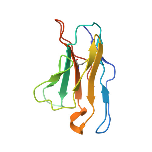
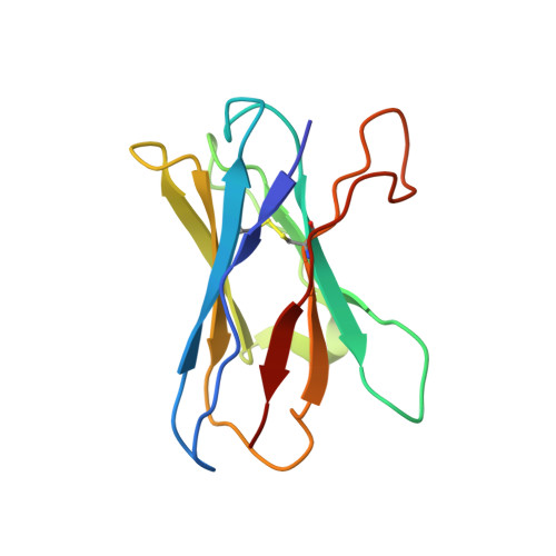
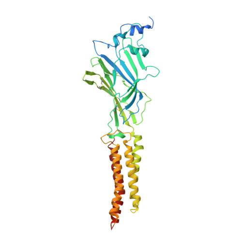
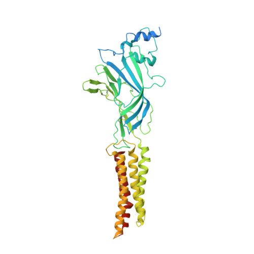
    Glycine receptors (GlyRs) are pentameric, 'Cys-loop' receptors that form chloride-permeable channels and mediate fast inhibitory signalling throughout the central nervous system 1,2 . In the spinal cord and brainstem, GlyRs regulate locomotion and cause movement disorders when mutated 2,3 . However, the stoichiometry of native GlyRs and the mechanism by which they are assembled remain unclear, despite extensive investigation 4-8 . Here we report cryo-electron microscopy structures of native GlyRs from pig spinal cord and brainstem, revealing structural insights into heteromeric receptors and their predominant subunit stoichiometry of 4α:1β. Within the heteromeric pentamer, the β(+)-α(-) interface adopts a structure that is distinct from the α(+)-α(-) and α(+)-β(-) interfaces. Furthermore, the β-subunit contains a unique phenylalanine residue that resides within the pore and disrupts the canonical picrotoxin site. These results explain why inclusion of the β-subunit breaks receptor symmetry and alters ion channel pharmacology. We also find incomplete receptor complexes and, by elucidating their structures, reveal the architectures of partially assembled α-trimers and α-tetramers.


  • Organizational Affiliation
    • Vollum Institute, Oregon Health and Science University, Portland, OR, USA.

Macromolecule Content 

  • Total Structure Weight: 369.52 kDa 
  • Atom Count: 20,982 
  • Modeled Residue Count: 2,558 
  • Deposited Residue Count: 3,185 
  • Unique protein chains: 4

Macromolecules

Find similar proteins by:|  3D Structure
Entity ID: 1
MoleculeChains  Sequence LengthOrganismDetailsImage
3D1 Fab Light ChainA [auth J],
C [auth K],
E [auth M],
G [auth I]
107Rattus norvegicusMutation(s): 0 
Find similar proteins by:|  3D Structure
Entity ID: 2
MoleculeChains  Sequence LengthOrganismDetailsImage
3D1 Fab Heavy ChainB [auth G],
D [auth F],
F [auth L],
H
118Rattus norvegicusMutation(s): 0 
Find similar proteins by:|  3D Structure
Entity ID: 3
MoleculeChains  Sequence LengthOrganismDetailsImage
Glycine receptor alpha 1I [auth D],
J [auth A],
K [auth B],
L [auth C]
447Sus scrofaMutation(s): 0 
Membrane Entity: Yes 
UniProt
Find proteins for A0A4X1TUM3 (Sus scrofa)
Explore A0A4X1TUM3 
Go to UniProtKB:  A0A4X1TUM3
Entity Groups
Sequence Clusters30% Identity50% Identity70% Identity90% Identity95% Identity100% Identity
UniProt GroupA0A4X1TUM3
Glycosylation
Glycosylation Sites: 1
Sequence Annotations
Expand
Reference Sequence
Find similar proteins by:|  3D Structure
Entity ID: 4
MoleculeChains  Sequence LengthOrganismDetailsImage
Glycine receptor betaM [auth E]497Sus scrofaMutation(s): 0 
Membrane Entity: Yes 
UniProt
Find proteins for Q6KBX4 (Sus scrofa)
Explore Q6KBX4 
Go to UniProtKB:  Q6KBX4
Entity Groups
Sequence Clusters30% Identity50% Identity70% Identity90% Identity95% Identity100% Identity
UniProt GroupQ6KBX4
Glycosylation
Glycosylation Sites: 1
Sequence Annotations
Expand
Reference Sequence

Oligosaccharides

Help  
Entity ID: 5
MoleculeChains Length2D Diagram GlycosylationD Interactions
alpha-D-mannopyranose-(1-2)-alpha-D-mannopyranose-(1-3)-beta-D-mannopyranose-(1-4)-2-acetamido-2-deoxy-beta-D-glucopyranose-(1-4)-2-acetamido-2-deoxy-beta-D-glucopyranoseN [auth T]5N-Glycosylation
Glycosylation Resources
GlyTouCan: G42227JK
GlyCosmos: G42227JK
GlyGen: G42227JK
Entity ID: 6
MoleculeChains Length2D Diagram GlycosylationD Interactions
alpha-D-mannopyranose-(1-3)-[alpha-D-mannopyranose-(1-6)]beta-D-mannopyranose-(1-4)-2-acetamido-2-deoxy-beta-D-glucopyranose-(1-4)-2-acetamido-2-deoxy-beta-D-glucopyranoseO [auth Z]5N-Glycosylation
Glycosylation Resources
GlyTouCan: G22768VO
GlyCosmos: G22768VO
GlyGen: G22768VO
Entity ID: 7
MoleculeChains Length2D Diagram GlycosylationD Interactions
alpha-D-mannopyranose-(1-6)-beta-D-mannopyranose-(1-4)-2-acetamido-2-deoxy-beta-D-glucopyranose-(1-4)-2-acetamido-2-deoxy-beta-D-glucopyranoseP [auth f],
Q [auth l]
4N-Glycosylation
Glycosylation Resources
GlyTouCan: G22573RC
GlyCosmos: G22573RC
GlyGen: G22573RC
Entity ID: 8
MoleculeChains Length2D Diagram GlycosylationD Interactions
beta-D-mannopyranose-(1-4)-2-acetamido-2-deoxy-beta-D-glucopyranose-(1-4)-2-acetamido-2-deoxy-beta-D-glucopyranoseR [auth r]3N-Glycosylation
Glycosylation Resources
GlyTouCan: G15407YE
GlyCosmos: G15407YE
GlyGen: G15407YE

Small Molecules

Ligands 11 Unique
IDChains Name / Formula / InChI Key2D Diagram3D Interactions
MYS

Query on MYS


Download:Ideal Coordinates CCD File
W [auth D]PENTADECANE
C15 H32
YCOZIPAWZNQLMR-UHFFFAOYSA-N
D12

Query on D12


Download:Ideal Coordinates CCD File
DA [auth A],
YA [auth C]
DODECANE
C12 H26
SNRUBQQJIBEYMU-UHFFFAOYSA-N
UND

Query on UND


Download:Ideal Coordinates CCD File
MB [auth E]UNDECANE
C11 H24
RSJKGSCJYJTIGS-UHFFFAOYSA-N
D10

Query on D10


Download:Ideal Coordinates CCD File
X [auth D]DECANE
C10 H22
DIOQZVSQGTUSAI-UHFFFAOYSA-N
DD9

Query on DD9


Download:Ideal Coordinates CCD File
MA [auth A],
PA [auth B],
U [auth D]
nonane
C9 H20
BKIMMITUMNQMOS-UHFFFAOYSA-N
OCT

Query on OCT


Download:Ideal Coordinates CCD File
AB [auth C],
HA [auth A],
IA [auth A],
LA [auth A]
N-OCTANE
C8 H18
TVMXDCGIABBOFY-UHFFFAOYSA-N
HP6

Query on HP6


Download:Ideal Coordinates CCD File
EA [auth A]
FA [auth A]
IB [auth E]
JB [auth E]
KB [auth E]
EA [auth A],
FA [auth A],
IB [auth E],
JB [auth E],
KB [auth E],
OA [auth B],
S [auth D],
T [auth D],
WA [auth B]
HEPTANE
C7 H16
IMNFDUFMRHMDMM-UHFFFAOYSA-N
HEX

Query on HEX


Download:Ideal Coordinates CCD File
GB [auth C],
KA [auth A],
RA [auth B]
HEXANE
C6 H14
VLKZOEOYAKHREP-UHFFFAOYSA-N
GLY

Query on GLY


Download:Ideal Coordinates CCD File
BA [auth A],
CA [auth A],
HB [auth E],
NA [auth B],
XA [auth C]
GLYCINE
C2 H5 N O2
DHMQDGOQFOQNFH-UHFFFAOYSA-N
LNK

Query on LNK


Download:Ideal Coordinates CCD File
BB [auth C]
GA [auth A]
JA [auth A]
LB [auth E]
NB [auth E]
BB [auth C],
GA [auth A],
JA [auth A],
LB [auth E],
NB [auth E],
QA [auth B],
SA [auth B],
V [auth D],
Y [auth D],
ZA [auth C]
PENTANE
C5 H12
OFBQJSOFQDEBGM-UHFFFAOYSA-N
NBU

Query on NBU


Download:Ideal Coordinates CCD File
AA [auth D]
CB [auth C]
DB [auth C]
EB [auth C]
FB [auth C]
AA [auth D],
CB [auth C],
DB [auth C],
EB [auth C],
FB [auth C],
TA [auth B],
UA [auth B],
VA [auth B],
Z [auth D]
N-BUTANE
C4 H10
IJDNQMDRQITEOD-UHFFFAOYSA-N

Experimental Data & Validation

Experimental Data

  • Method: ELECTRON MICROSCOPY
  • Resolution: 2.70 Å
  • Aggregation State: PARTICLE 
  • Reconstruction Method: SINGLE PARTICLE 
EM Software:
TaskSoftware PackageVersion
RECONSTRUCTIONcryoSPARC

Structure Validation

View Full Validation Report


Entry History 

& Funding Information

Deposition Data


Funding OrganizationLocationGrant Number
National Institutes of Health/National Institute of General Medical Sciences (NIH/NIGMS)United StatesR01 GM100400

Revision History  (Full details and data files)

  • Version 1.0: 2021-09-29
    Type: Initial release
  • Version 1.1: 2021-10-06
    Changes: Database references
  • Version 1.2: 2021-12-01
    Changes: Database references
  • Version 1.3: 2024-10-23
    Changes: Data collection, Structure summary