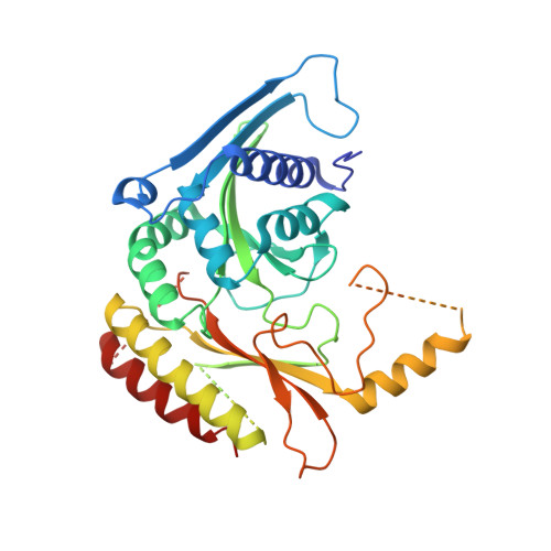
7N81

Crystal Structure of PI5P4KIIBeta complex with CC260


Experimental Data Snapshot

  • Method: X-RAY DIFFRACTION
  • Resolution: 2.70 Å
  • R-Value Free: 0.281 
  • R-Value Work: 0.187 
  • R-Value Observed: 0.192 

Starting Model: experimental
View more details

wwPDB Validation   3D Report Full Report


Ligand Structure Quality Assessment 


This is version 1.3 of the entry. See complete history


Literature

Pharmacological inhibition of PI5P4K alpha / beta disrupts cell energy metabolism and selectively kills p53-null tumor cells.

Chen, S.Chandra Tjin, C.Gao, X.Xue, Y.Jiao, H.Zhang, R.Wu, M.He, Z.Ellman, J.Ha, Y.

(2021) Proc Natl Acad Sci U S A 118

  • DOI: https://doi.org/10.1073/pnas.2002486118
  • Primary Citation of Related Structures:  
    7N6Z, 7N71, 7N7J, 7N7K, 7N7L, 7N7M, 7N7N, 7N7O, 7N80, 7N81

  • PubMed Abstract: 

    Most human cancer cells harbor loss-of-function mutations in the p53 tumor suppressor gene. Genetic experiments have shown that phosphatidylinositol 5-phosphate 4-kinase α and β (PI5P4Kα and PI5P4Kβ) are essential for the development of late-onset tumors in mice with germline p53 deletion, but the mechanism underlying this acquired dependence remains unclear. PI5P4K has been previously implicated in metabolic regulation. Here, we show that inhibition of PI5P4Kα/β kinase activity by a potent and selective small-molecule probe disrupts cell energy homeostasis, causing AMPK activation and mTORC1 inhibition in a variety of cell types. Feedback through the S6K/insulin receptor substrate (IRS) loop contributes to insulin hypersensitivity and enhanced PI3K signaling in terminally differentiated myotubes. Most significantly, the energy stress induced by PI5P4Kαβ inhibition is selectively toxic toward p53-null tumor cells. The chemical probe, and the structural basis for its exquisite specificity, provide a promising platform for further development, which may lead to a novel class of diabetes and cancer drugs.


  • Organizational Affiliation

    Department of Pharmacology, Yale School of Medicine, New Haven, CT 06520.


Macromolecules
Find similar proteins by:  (by identity cutoff)  |  3D Structure
Entity ID: 1
MoleculeChains Sequence LengthOrganismDetailsImage
Phosphatidylinositol 5-phosphate 4-kinase type-2 beta
A, B
392Homo sapiensMutation(s): 0 
Gene Names: PIP4K2BPIP5K2B
EC: 2.7.1.149
UniProt & NIH Common Fund Data Resources
Find proteins for P78356 (Homo sapiens)
Go to UniProtKB:  P78356
PHAROS:  P78356
GTEx:  ENSG00000276293 
Sequence Annotations
Expand
  • Reference Sequence
Small Molecules
Ligands 1 Unique
IDChains Name / Formula / InChI Key2D Diagram3D Interactions
HKP (Subject of Investigation/LOI)
Query on HKP

Download Ideal Coordinates CCD File 
C [auth B](7R)-8-cyclopentyl-7-(cyclopentylmethyl)-2-[(3,5-dichloro-4-hydroxyphenyl)amino]-5-methyl-7,8-dihydropteridin-6(5H)-one
C24 H29 Cl2 N5 O2
AEZJZTFWCDAUDF-LJQANCHMSA-N
Binding Affinity Annotations 
IDSourceBinding Affinity
HKP BindingDB:  7N81 IC50: 30 (nM) from 1 assay(s)
Experimental Data & Validation

Experimental Data

  • Method: X-RAY DIFFRACTION
  • Resolution: 2.70 Å
  • R-Value Free: 0.281 
  • R-Value Work: 0.187 
  • R-Value Observed: 0.192 
  • Space Group: P 1 21 1
Unit Cell:
Length ( Å )Angle ( ˚ )
a = 73.523α = 90
b = 51.748β = 95.38
c = 107.606γ = 90
Software Package:
Software NamePurpose
HKL-2000data scaling
REFMACrefinement
PDB_EXTRACTdata extraction
HKL-2000data reduction
REFMACphasing

Structure Validation

View Full Validation Report


Ligand Structure Quality Assessment 


Entry History 

Deposition Data

Revision History  (Full details and data files)

  • Version 1.0: 2021-06-30
    Type: Initial release
  • Version 1.1: 2021-07-14
    Changes: Database references
  • Version 1.2: 2023-10-04
    Changes: Data collection, Database references
  • Version 1.3: 2023-10-18
    Changes: Refinement description