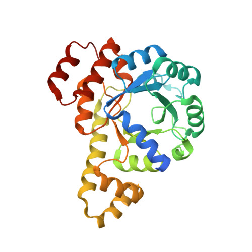
7NHE

Crystal structure of Arabidopsis thaliana Pdx1K166R-I333 complex


Experimental Data Snapshot

  • Method: X-RAY DIFFRACTION
  • Resolution: 2.23 Å
  • R-Value Free: 0.219 
  • R-Value Work: 0.175 
  • R-Value Observed: 0.177 

Starting Model: experimental
View more details

wwPDB Validation   3D Report Full Report


Ligand Structure Quality Assessment 


This is version 1.3 of the entry. See complete history


Literature

Trapping and structural characterisation of a covalent intermediate in vitamin B 6 biosynthesis catalysed by the Pdx1 PLP synthase.

Rodrigues, M.J.Giri, N.Royant, A.Zhang, Y.Bolton, R.Evans, G.Ealick, S.E.Begley, T.Tews, I.

(2022) RSC Chem Biol 3: 227-230

  • DOI: https://doi.org/10.1039/d1cb00160d
  • Primary Citation of Related Structures:  
    7NHE, 7NHF

  • PubMed Abstract: 

    The Pdx1 enzyme catalyses condensation of two carbohydrates and ammonia to form pyridoxal 5-phosphate (PLP) via an imine relay mechanism of carbonyl intermediates. The I 333 intermediate characterised here using structural, UV-vis absorption spectroscopy and mass spectrometry analyses rationalises stereoselective deprotonation and subsequent substrate assisted phosphate elimination, central to PLP biosynthesis.


  • Organizational Affiliation

    Biological Sciences, Institute for Life Sciences, University of Southampton Southampton SO17 1BJ UK ivo.tews@soton.ac.uk.


Macromolecules
Find similar proteins by:  (by identity cutoff)  |  3D Structure
Entity ID: 1
MoleculeChains Sequence LengthOrganismDetailsImage
Pyridoxal 5'-phosphate synthase subunit PDX1.3
A, B, C, D
291Arabidopsis thalianaMutation(s): 1 
Gene Names: PDX13GIP2PDX1L3RSR4At5g01410T10O8.120
EC: 4.3.3.6
UniProt
Find proteins for Q8L940 (Arabidopsis thaliana)
Explore Q8L940 
Go to UniProtKB:  Q8L940
Entity Groups  
Sequence Clusters30% Identity50% Identity70% Identity90% Identity95% Identity100% Identity
UniProt GroupQ8L940
Sequence Annotations
Expand
  • Reference Sequence
Small Molecules
Ligands 2 Unique
IDChains Name / Formula / InChI Key2D Diagram3D Interactions
KPR (Subject of Investigation/LOI)
Query on KPR

Download Ideal Coordinates CCD File 
F [auth A],
H [auth B],
J [auth C],
L [auth D]
[(~{E},4~{S})-4-azanyl-3-oxidanylidene-pent-1-enyl] dihydrogen phosphate
C5 H10 N O5 P
RTVKPJRIOVZREZ-ZPYNKGFJSA-N
PO4
Query on PO4

Download Ideal Coordinates CCD File 
E [auth A],
G [auth B],
I [auth C],
K [auth D]
PHOSPHATE ION
O4 P
NBIIXXVUZAFLBC-UHFFFAOYSA-K
Experimental Data & Validation

Experimental Data

  • Method: X-RAY DIFFRACTION
  • Resolution: 2.23 Å
  • R-Value Free: 0.219 
  • R-Value Work: 0.175 
  • R-Value Observed: 0.177 
  • Space Group: H 3
Unit Cell:
Length ( Å )Angle ( ˚ )
a = 177.973α = 90
b = 177.973β = 90
c = 115.121γ = 120
Software Package:
Software NamePurpose
PHENIXrefinement
Aimlessdata scaling
PDB_EXTRACTdata extraction
MOLREPphasing
Cootmodel building
DIALSdata reduction

Structure Validation

View Full Validation Report


Ligand Structure Quality Assessment 


Entry History 

Deposition Data

Revision History  (Full details and data files)

  • Version 1.0: 2021-12-22
    Type: Initial release
  • Version 1.1: 2022-04-13
    Changes: Database references
  • Version 1.2: 2024-01-31
    Changes: Data collection, Refinement description
  • Version 1.3: 2024-10-16
    Changes: Structure summary