7P3S | pdb_00007p3s

Crystal structure of Schistosoma mansoni HDAC8 complexed with a benzohydroxamate inhibitor 12


Experimental Data Snapshot

  • Method: X-RAY DIFFRACTION
  • Resolution: 1.55 Å
  • R-Value Free: 
    0.193 (Depositor), 0.195 (DCC) 
  • R-Value Work: 
    0.169 (Depositor), 0.170 (DCC) 
  • R-Value Observed: 
    0.170 (Depositor) 

Starting Model: experimental
View more details

wwPDB Validation 3D Report Full Report

Validation slider image for 7P3S

Ligand Structure Quality Assessment 


This is version 1.1 of the entry. See complete history

Literature

Synthesis, structure-activity relationships, cocrystallization and cellular characterization of novel smHDAC8 inhibitors for the treatment of schistosomiasis.

Ghazy, E.Heimburg, T.Lancelot, J.Zeyen, P.Schmidtkunz, K.Truhn, A.Darwish, S.Simoben, C.V.Shaik, T.B.Erdmann, F.Schmidt, M.Robaa, D.Romier, C.Jung, M.Pierce, R.Sippl, W.

(2021) Eur J Med Chem 225: 113745-113745

  • DOI: https://doi.org/10.1016/j.ejmech.2021.113745
  • Primary Citation Related Structures: 
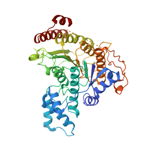
    7P3S

  • PubMed Abstract: 

    Schistosomiasis is a major neglected parasitic disease that affects more than 265 million people worldwide and for which the control strategy consists of mass treatment with the only available drug, praziquantel. In this study, we chemically optimized our previously reported benzhydroxamate-based inhibitors of Schistosoma mansoni histone deacetylase 8 (smHDAC8). Crystallographic analysis provided insights into the inhibition mode of smHDAC8 activity by the highly potent inhibitor 5o. Structure-based optimization of the novel inhibitors was carried out using the available crystal structures as well as docking studies on smHDAC8. The compounds were evaluated in screens for inhibitory activity against schistosome and human HDACs (hHDAC). The in vitro and docking results were used for detailed structure activity relationships. The synthesized compounds were further investigated for their lethality against the schistosome larval stage using a fluorescence-based assay. The most promising inhibitor 5o showed significant dose-dependent killing of the schistosome larvae and markedly impaired egg laying of adult worm pairs maintained in culture.


  • Organizational Affiliation
    • Institute of Pharmacy, Martin-Luther University of Halle-Wittenberg, 06120, Halle/Saale, Germany.

Macromolecule Content 

  • Total Structure Weight: 205.15 kDa 
  • Atom Count: 14,397 
  • Modeled Residue Count: 1,620 
  • Deposited Residue Count: 1,788 
  • Unique protein chains: 1

Macromolecules

Find similar proteins by:|  3D Structure
Entity ID: 1
MoleculeChains  Sequence LengthOrganismDetailsImage
Histone deacetylase
A, B, C, D
447Schistosoma mansoniMutation(s): 0 
Gene Names: HDAC8
EC: 3.5.1.98
UniProt
Find proteins for A5H660 (Schistosoma mansoni)
Explore A5H660 
Go to UniProtKB:  A5H660
Entity Groups
Sequence Clusters30% Identity50% Identity70% Identity90% Identity95% Identity100% Identity
UniProt GroupA5H660
Sequence Annotations
Expand
Reference Sequence

Small Molecules

Ligands 4 Unique
IDChains Name / Formula / InChI Key2D Diagram3D Interactions
5IB
(Subject of Investigation/LOI)

Query on 5IB


Download:Ideal Coordinates CCD File
K [auth A],
L [auth B],
Q [auth C],
W [auth D]
~{N}-[2-methoxy-5-(oxidanylcarbamoyl)phenyl]dibenzofuran-4-carboxamide
C21 H16 N2 O5
RFROQVRGDKTHLN-UHFFFAOYSA-N
GOL

Query on GOL


Download:Ideal Coordinates CCD File
AA [auth D]
BA [auth D]
H [auth A]
I [auth A]
J [auth A]
AA [auth D],
BA [auth D],
H [auth A],
I [auth A],
J [auth A],
P [auth B],
U [auth C],
V [auth C]
GLYCEROL
C3 H8 O3
PEDCQBHIVMGVHV-UHFFFAOYSA-N
ZN

Query on ZN


Download:Ideal Coordinates CCD File
E [auth A],
M [auth B],
R [auth C],
X [auth D]
ZINC ION
Zn
PTFCDOFLOPIGGS-UHFFFAOYSA-N
K

Query on K


Download:Ideal Coordinates CCD File
F [auth A]
G [auth A]
N [auth B]
O [auth B]
S [auth C]
F [auth A],
G [auth A],
N [auth B],
O [auth B],
S [auth C],
T [auth C],
Y [auth D],
Z [auth D]
POTASSIUM ION
K
NPYPAHLBTDXSSS-UHFFFAOYSA-N
Binding Affinity Annotations 
IDSourceBinding Affinity
5IB BindingDB:  7P3S IC50: 270 (nM) from 1 assay(s)

Experimental Data & Validation

Experimental Data

  • Method: X-RAY DIFFRACTION
  • Resolution: 1.55 Å
  • R-Value Free:  0.193 (Depositor), 0.195 (DCC) 
  • R-Value Work:  0.169 (Depositor), 0.170 (DCC) 
  • R-Value Observed: 0.170 (Depositor) 
Space Group: P 1
Unit Cell:
Length ( Å )Angle ( ˚ )
a = 70.39α = 78.05
b = 70.39β = 75.57
c = 97.94γ = 85.69
Software Package:
Software NamePurpose
PHENIXrefinement
PDB_EXTRACTdata extraction
XDSdata reduction
XDSdata scaling
PHENIXphasing

Structure Validation

View Full Validation Report


Ligand Structure Quality Assessment 


Entry History 

& Funding Information

Deposition Data


Funding OrganizationLocationGrant Number
European Communitys Seventh Framework ProgrammeEuropean Union241865
European Communitys Seventh Framework ProgrammeEuropean Union602080

Revision History  (Full details and data files)

  • Version 1.0: 2021-09-15
    Type: Initial release
  • Version 1.1: 2024-01-31
    Changes: Data collection, Database references, Refinement description