7Q24 | pdb_00007q24

Crystal structure of Angiotensin-1 converting enzyme N-domain in complex with dual ACE/NEP inhibitor AD011


Experimental Data Snapshot

  • Method: X-RAY DIFFRACTION
  • Resolution: 2.00 Å
  • R-Value Free: 
    0.214 (Depositor), 0.213 (DCC) 
  • R-Value Work: 
    0.185 (Depositor), 0.183 (DCC) 
  • R-Value Observed: 
    0.186 (Depositor) 

Starting Model: experimental
View more details

wwPDB Validation 3D Report Full Report

Validation slider image for 7Q24

Ligand Structure Quality Assessment 


This is version 1.3 of the entry. See complete history

Literature

Probing the Requirements for Dual Angiotensin-Converting Enzyme C-Domain Selective/Neprilysin Inhibition.

Arendse, L.B.Cozier, G.E.Eyermann, C.J.Basarab, G.S.Schwager, S.L.Chibale, K.Acharya, K.R.Sturrock, E.D.

(2022) J Med Chem 65: 3371-3387

  • DOI: https://doi.org/10.1021/acs.jmedchem.1c01924
  • Primary Citation Related Structures: 
    7Q24, 7Q25, 7Q26, 7Q27, 7Q28, 7Q29

  • PubMed Abstract: 

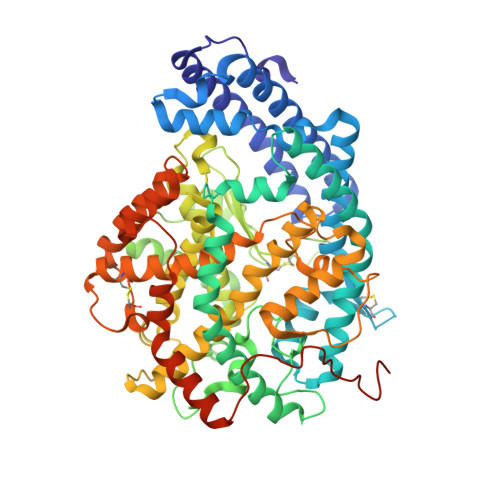
    Selective inhibition of the angiotensin-converting enzyme C-domain (cACE) and neprilysin (NEP), leaving the ACE N-domain (nACE) free to degrade bradykinin and other peptides, has the potential to provide the potent antihypertensive and cardioprotective benefits observed for nonselective dual ACE/NEP inhibitors, such as omapatrilat, without the increased risk of adverse effects. We have synthesized three 1-carboxy-3-phenylpropyl dipeptide inhibitors with nanomolar potency based on the previously reported C-domain selective ACE inhibitor lisinopril-tryptophan (LisW) to probe the structural requirements for potent dual cACE/NEP inhibition. Here we report the synthesis, enzyme kinetic data, and high-resolution crystal structures of these inhibitors bound to nACE and cACE, providing valuable insight into the factors driving potency and selectivity. Overall, these results highlight the importance of the interplay between the S 1 ' and S 2 ' subsites for ACE domain selectivity, providing guidance for future chemistry efforts toward the development of dual cACE/NEP inhibitors.


  • Organizational Affiliation
    • Institute of Infectious Disease and Molecular Medicine, University of Cape Town, Observatory, Cape Town 7925, South Africa.

Macromolecule Content 

  • Total Structure Weight: 151.12 kDa 
  • Atom Count: 10,964 
  • Modeled Residue Count: 1,215 
  • Deposited Residue Count: 1,258 
  • Unique protein chains: 1

Macromolecules

Find similar proteins by:|  3D Structure
Entity ID: 1
MoleculeChains  Sequence LengthOrganismDetailsImage
Angiotensin-converting enzyme
A, B
629Homo sapiensMutation(s): 8 
Gene Names: ACEDCPDCP1
EC: 3.2.1 (PDB Primary Data), 3.4.15.1 (PDB Primary Data)
UniProt & NIH Common Fund Data Resources
Find proteins for P12821 (Homo sapiens)
Explore P12821 
Go to UniProtKB:  P12821
PHAROS:  P12821
GTEx:  ENSG00000159640 
Entity Groups
Sequence Clusters30% Identity50% Identity70% Identity90% Identity95% Identity100% Identity
UniProt GroupP12821
Glycosylation
Glycosylation Sites: 3Go to GlyGen: P12821-1
Sequence Annotations
Expand
Reference Sequence

Oligosaccharides

Help  
Entity ID: 2
MoleculeChains Length2D Diagram GlycosylationD Interactions
2-acetamido-2-deoxy-beta-D-glucopyranose-(1-4)-2-acetamido-2-deoxy-beta-D-glucopyranose
C
2N-Glycosylation
Glycosylation Resources
GlyTouCan: G42666HT
GlyCosmos: G42666HT
GlyGen: G42666HT
Entity ID: 3
MoleculeChains Length2D Diagram GlycosylationD Interactions
beta-D-mannopyranose-(1-4)-2-acetamido-2-deoxy-beta-D-glucopyranose-(1-4)-[alpha-L-fucopyranose-(1-6)]2-acetamido-2-deoxy-beta-D-glucopyranose
D, F
4N-Glycosylation
Glycosylation Resources
GlyTouCan: G32152BH
GlyCosmos: G32152BH
GlyGen: G32152BH
Entity ID: 4
MoleculeChains Length2D Diagram GlycosylationD Interactions
alpha-L-fucopyranose-(1-6)-2-acetamido-2-deoxy-beta-D-glucopyranose
E, G
2N-Glycosylation
Glycosylation Resources
GlyTouCan: G86851RC
GlyCosmos: G86851RC
GlyGen: G86851RC

Small Molecules

Ligands 11 Unique
IDChains Name / Formula / InChI Key2D Diagram3D Interactions
8KC
(Subject of Investigation/LOI)

Query on 8KC


Download:Ideal Coordinates CCD File
EA [auth B],
S [auth A]
(2~{S})-2-[[(2~{S})-1-[[(2~{S})-3-(1~{H}-indol-3-yl)-1-oxidanyl-1-oxidanylidene-propan-2-yl]amino]-1-oxidanylidene-hexan-2-yl]amino]-4-phenyl-butanoic acid
C27 H33 N3 O5
YWBHCYRCVYEOPE-HJOGWXRNSA-N
P33

Query on P33


Download:Ideal Coordinates CCD File
J [auth A]3,6,9,12,15,18-HEXAOXAICOSANE-1,20-DIOL
C14 H30 O8
XPJRQAIZZQMSCM-UHFFFAOYSA-N
P6G

Query on P6G


Download:Ideal Coordinates CCD File
N [auth A]HEXAETHYLENE GLYCOL
C12 H26 O7
IIRDTKBZINWQAW-UHFFFAOYSA-N
NAG

Query on NAG


Download:Ideal Coordinates CCD File
U [auth B]2-acetamido-2-deoxy-beta-D-glucopyranose
C8 H15 N O6
OVRNDRQMDRJTHS-FMDGEEDCSA-N
PG4

Query on PG4


Download:Ideal Coordinates CCD File
T [auth B],
W [auth B]
TETRAETHYLENE GLYCOL
C8 H18 O5
UWHCKJMYHZGTIT-UHFFFAOYSA-N
PEG

Query on PEG


Download:Ideal Coordinates CCD File
AA [auth B],
H [auth A],
I [auth A],
K [auth A]
DI(HYDROXYETHYL)ETHER
C4 H10 O3
MTHSVFCYNBDYFN-UHFFFAOYSA-N
ZN

Query on ZN


Download:Ideal Coordinates CCD File
BA [auth B],
P [auth A]
ZINC ION
Zn
PTFCDOFLOPIGGS-UHFFFAOYSA-N
EDO

Query on EDO


Download:Ideal Coordinates CCD File
M [auth A],
O [auth A],
V [auth B],
Y [auth B]
1,2-ETHANEDIOL
C2 H6 O2
LYCAIKOWRPUZTN-UHFFFAOYSA-N
ACT

Query on ACT


Download:Ideal Coordinates CCD File
L [auth A],
X [auth B],
Z [auth B]
ACETATE ION
C2 H3 O2
QTBSBXVTEAMEQO-UHFFFAOYSA-M
CL

Query on CL


Download:Ideal Coordinates CCD File
CA [auth B],
Q [auth A]
CHLORIDE ION
Cl
VEXZGXHMUGYJMC-UHFFFAOYSA-M
MG

Query on MG


Download:Ideal Coordinates CCD File
DA [auth B],
R [auth A]
MAGNESIUM ION
Mg
JLVVSXFLKOJNIY-UHFFFAOYSA-N
Binding Affinity Annotations 
IDSourceBinding Affinity
8KC BindingDB:  7Q24 Ki: min: 100, max: 3790 (nM) from 2 assay(s)

Experimental Data & Validation

Experimental Data

  • Method: X-RAY DIFFRACTION
  • Resolution: 2.00 Å
  • R-Value Free:  0.214 (Depositor), 0.213 (DCC) 
  • R-Value Work:  0.185 (Depositor), 0.183 (DCC) 
  • R-Value Observed: 0.186 (Depositor) 
Space Group: P 1
Unit Cell:
Length ( Å )Angle ( ˚ )
a = 72.766α = 88.931
b = 77.608β = 64.553
c = 81.647γ = 75.009
Software Package:
Software NamePurpose
PHENIXrefinement
DIALSdata reduction
Aimlessdata scaling
PHASERphasing

Structure Validation

View Full Validation Report


Ligand Structure Quality Assessment 


Entry History 

& Funding Information

Deposition Data


Funding OrganizationLocationGrant Number
Medical Research Council (MRC, United Kingdom)United KingdomMR/M026647/1

Revision History  (Full details and data files)

  • Version 1.0: 2022-02-16
    Type: Initial release
  • Version 1.1: 2022-03-09
    Changes: Database references
  • Version 1.2: 2024-01-31
    Changes: Data collection, Refinement description
  • Version 1.3: 2024-11-06
    Changes: Structure summary