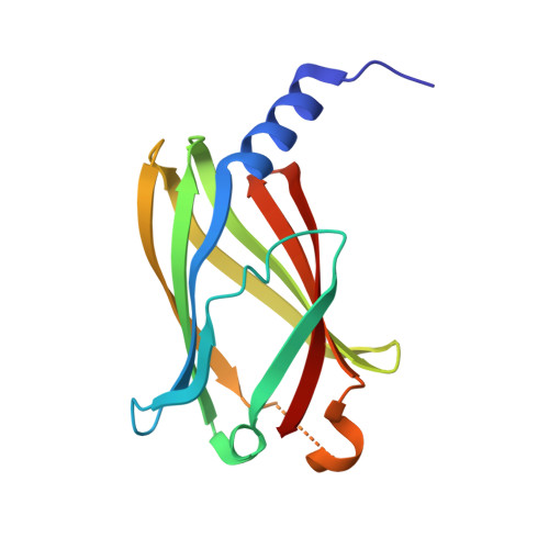
7Q9Q | pdb_00007q9q

Crystal structure of PDE6D Geranylgeranylated cystein complex


Experimental Data Snapshot

  • Method: X-RAY DIFFRACTION
  • Resolution: 1.45 Å
  • R-Value Free: 
    0.274 (Depositor), 0.280 (DCC) 
  • R-Value Work: 
    0.225 (Depositor), 0.230 (DCC) 

Starting Model: experimental
View more details

wwPDB Validation   3D Report Full Report


Ligand Structure Quality Assessment 

Created with Raphaël 2.3.0Worse 01 BetterLigand structure goodness of fit to experimental dataBest fitted GERClick on this verticalbar to view detailsBest fitted CMTClick on this verticalbar to view details

This is version 1.2 of the entry. See complete history


Literature

Stabilization of the RAS:PDE6D Complex Is a Novel Strategy to Inhibit RAS Signaling.

Yelland, T.Garcia, E.Parry, C.Kowalczyk, D.Wojnowska, M.Gohlke, A.Zalar, M.Cameron, K.Goodwin, G.Yu, Q.Zhu, P.C.ElMaghloob, Y.Pugliese, A.Archibald, L.Jamieson, A.Chen, Y.X.McArthur, D.Bower, J.Ismail, S.

(2022) J Med Chem 65: 1898-1914

  • DOI: https://doi.org/10.1021/acs.jmedchem.1c01265
  • Primary Citation of Related Structures:  
    7Q9Q, 7Q9R, 7Q9S, 7Q9U, 7QF9, 7QJK

  • PubMed Abstract: 

    RAS is a major anticancer drug target which requires membrane localization to activate downstream signal transduction. The direct inhibition of RAS has proven to be challenging. Here, we present a novel strategy for targeting RAS by stabilizing its interaction with the prenyl-binding protein PDE6D and disrupting its localization. Using rationally designed RAS point mutations, we were able to stabilize the RAS:PDE6D complex by increasing the affinity of RAS for PDE6D, which resulted in the redirection of RAS to the cytoplasm and the primary cilium and inhibition of oncogenic RAS/ERK signaling. We developed an SPR fragment screening and identified fragments that bind at the KRAS:PDE6D interface, as shown through cocrystal structures. Finally, we show that the stoichiometric ratios of KRAS:PDE6D vary in different cell lines, suggesting that the impact of this strategy might be cell-type-dependent. This study forms the foundation from which a potential anticancer small-molecule RAS:PDE6D complex stabilizer could be developed.


  • Organizational Affiliation

    CRUK Beatson Institute, Glasgow G61 1BD, United Kingdom.


Macromolecules
Find similar proteins by:  (by identity cutoff)  |  3D Structure
Entity ID: 1
MoleculeChains Sequence LengthOrganismDetailsImage
Retinal rod rhodopsin-sensitive cGMP 3',5'-cyclic phosphodiesterase subunit deltaA [auth BBB]153Homo sapiensMutation(s): 0 
Gene Names: PDE6DPDED
UniProt & NIH Common Fund Data Resources
Find proteins for O43924 (Homo sapiens)
Explore O43924 
Go to UniProtKB:  O43924
PHAROS:  O43924
GTEx:  ENSG00000156973 
Entity Groups  
Sequence Clusters30% Identity50% Identity70% Identity90% Identity95% Identity100% Identity
UniProt GroupO43924
Sequence Annotations
Expand
  • Reference Sequence
Experimental Data & Validation

Experimental Data

  • Method: X-RAY DIFFRACTION
  • Resolution: 1.45 Å
  • R-Value Free:  0.274 (Depositor), 0.280 (DCC) 
  • R-Value Work:  0.225 (Depositor), 0.230 (DCC) 
Space Group: P 1
Unit Cell:
Length ( Å )Angle ( ˚ )
a = 32.048α = 77.725
b = 35.289β = 68.98
c = 37.645γ = 69.228
Software Package:
Software NamePurpose
REFMACrefinement
XDSdata reduction
Aimlessdata scaling
PHASERphasing

Structure Validation

View Full Validation Report


Ligand Structure Quality Assessment 

Created with Raphaël 2.3.0Worse 01 BetterLigand structure goodness of fit to experimental dataBest fitted GERClick on this verticalbar to view detailsBest fitted CMTClick on this verticalbar to view details

Entry History & Funding Information

Deposition Data


Funding OrganizationLocationGrant Number
Cancer Research UKUnited Kingdom--

Revision History  (Full details and data files)

  • Version 1.0: 2022-01-19
    Type: Initial release
  • Version 1.1: 2022-06-01
    Changes: Database references
  • Version 1.2: 2024-01-31
    Changes: Data collection, Derived calculations, Refinement description