7R63 | pdb_00007r63

Nb82, a nanobody against voltage gated sodium channels Nav1.4 and Nav1.5


Experimental Data Snapshot

  • Method: X-RAY DIFFRACTION
  • Resolution: 2.00 Å
  • R-Value Free: 
    0.244 (Depositor), 0.250 (DCC) 
  • R-Value Work: 
    0.184 (Depositor) 
  • R-Value Observed: 
    0.187 (Depositor) 

Starting Model: experimental
View more details

wwPDB Validation 3D Report Full Report

Validation slider image for 7R63

This is version 1.2 of the entry. See complete history

Literature

Development of high-affinity nanobodies specific for Na V 1.4 and Na V 1.5 voltage-gated sodium channel isoforms.

Srinivasan, L.Alzogaray, V.Selvakumar, D.Nathan, S.Yoder, J.B.Wright, K.M.Klinke, S.Nwafor, J.N.Labanda, M.S.Goldbaum, F.A.Schon, A.Freire, E.Tomaselli, G.F.Amzel, L.M.Ben-Johny, M.Gabelli, S.B.

(2022) J Biological Chem 298: 101763-101763

  • DOI: https://doi.org/10.1016/j.jbc.2022.101763
  • Primary Citation Related Structures: 
    7R63

  • PubMed Abstract: 

    Voltage-gated sodium channels, Na V s, are responsible for the rapid rise of action potentials in excitable tissues. Na V channel mutations have been implicated in several human genetic diseases, such as hypokalemic periodic paralysis, myotonia, and long-QT and Brugada syndromes. Here, we generated high-affinity anti-Na V nanobodies (Nbs), Nb17 and Nb82, that recognize the Na V 1.4 (skeletal muscle) and Na V 1.5 (cardiac muscle) channel isoforms. These Nbs were raised in llama (Lama glama) and selected from a phage display library for high affinity to the C-terminal (CT) region of Na V 1.4. The Nbs were expressed in Escherichia coli, purified, and biophysically characterized. Development of high-affinity Nbs specifically targeting a given human Na V isoform has been challenging because they usually show undesired crossreactivity for different Na V isoforms. Our results show, however, that Nb17 and Nb82 recognize the CTNa V 1.4 or CTNa V 1.5 over other CTNav isoforms. Kinetic experiments by biolayer interferometry determined that Nb17 and Nb82 bind to the CTNa V 1.4 and CTNa V 1.5 with high affinity (K D ∼ 40-60 nM). In addition, as proof of concept, we show that Nb82 could detect Na V 1.4 and Na V 1.5 channels in mammalian cells and tissues by Western blot. Furthermore, human embryonic kidney cells expressing holo Na V 1.5 channels demonstrated a robust FRET-binding efficiency for Nb17 and Nb82. Our work lays the foundation for developing Nbs as anti-Na V reagents to capture Na V s from cell lysates and as molecular visualization agents for Na V s.


  • Organizational Affiliation
    • Department of Biophysics and Biophysical Chemistry, The Johns Hopkins School of Medicine, Baltimore, Maryland, USA.

Macromolecule Content 

  • Total Structure Weight: 62.14 kDa 
  • Atom Count: 4,363 
  • Modeled Residue Count: 513 
  • Deposited Residue Count: 548 
  • Unique protein chains: 1

Macromolecules

Find similar proteins by:|  3D Structure
Entity ID: 1
MoleculeChains  Sequence LengthOrganismDetailsImage
Nanobody Nb82
A, B, C, D
137Lama glamaMutation(s): 0 
Gene Names: VHH

Small Molecules

Ligands 4 Unique
IDChains Name / Formula / InChI Key2D Diagram3D Interactions
PG0

Query on PG0


Download:Ideal Coordinates CCD File
H [auth A],
M [auth B],
T [auth D]
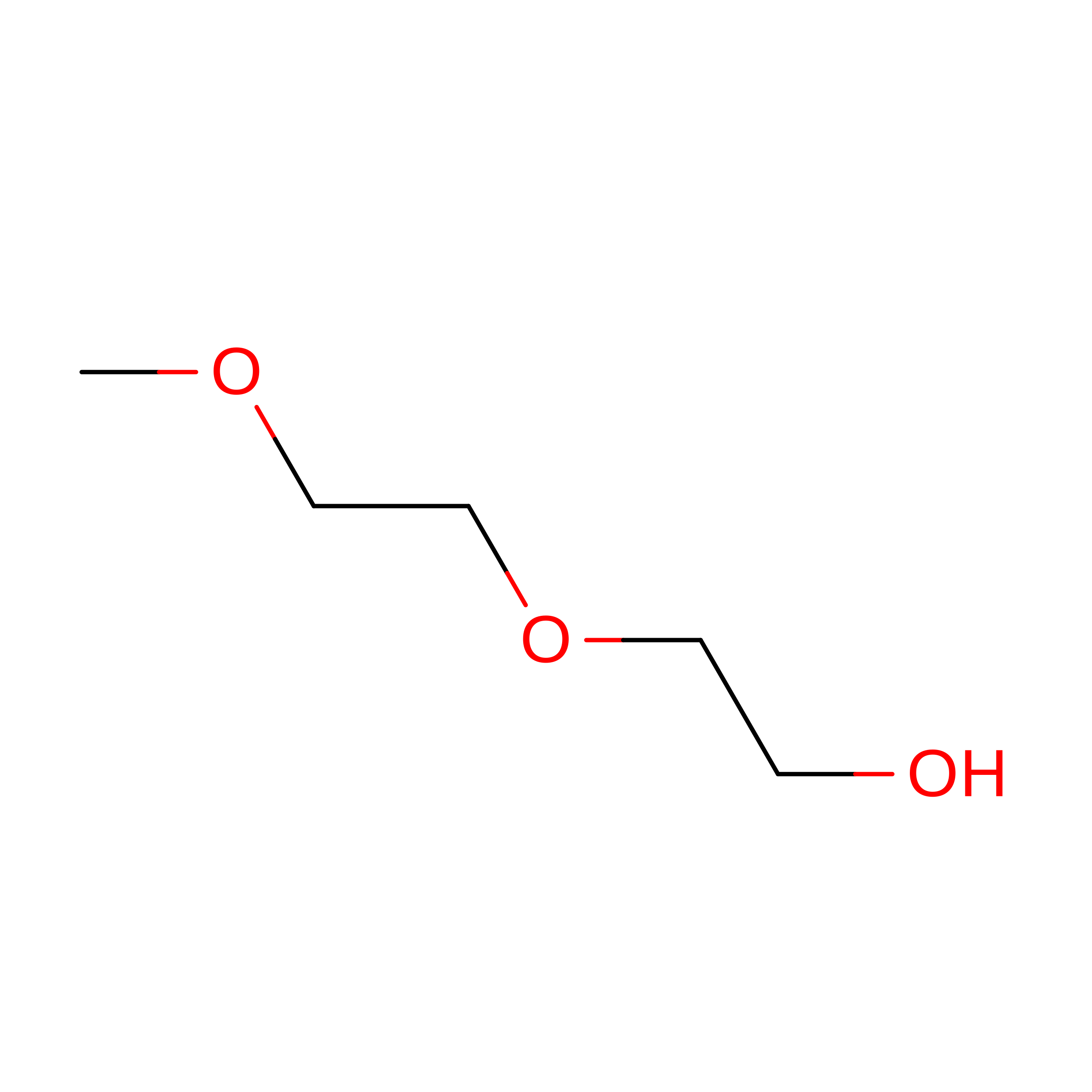
2-(2-METHOXYETHOXY)ETHANOL
C5 H12 O3
SBASXUCJHJRPEV-UHFFFAOYSA-N
SO4

Query on SO4


Download:Ideal Coordinates CCD File
E [auth A]
F [auth A]
G [auth A]
K [auth B]
L [auth B]
E [auth A],
F [auth A],
G [auth A],
K [auth B],
L [auth B],
O [auth C],
R [auth D],
S [auth D]
SULFATE ION
O4 S
QAOWNCQODCNURD-UHFFFAOYSA-L
CL

Query on CL


Download:Ideal Coordinates CCD File
P [auth C]CHLORIDE ION
Cl
VEXZGXHMUGYJMC-UHFFFAOYSA-M
NA

Query on NA


Download:Ideal Coordinates CCD File
I [auth B],
J [auth B],
N [auth C],
Q [auth D]
SODIUM ION
Na
FKNQFGJONOIPTF-UHFFFAOYSA-N

Experimental Data & Validation

Experimental Data

  • Method: X-RAY DIFFRACTION
  • Resolution: 2.00 Å
  • R-Value Free:  0.244 (Depositor), 0.250 (DCC) 
  • R-Value Work:  0.184 (Depositor) 
  • R-Value Observed: 0.187 (Depositor) 
Space Group: C 2 2 21
Unit Cell:
Length ( Å )Angle ( ˚ )
a = 77.745α = 90
b = 82.474β = 90
c = 170.7γ = 90
Software Package:
Software NamePurpose
Aimlessdata scaling
REFMACrefinement
PDB_EXTRACTdata extraction
XDSdata reduction
PHASERphasing

Structure Validation

View Full Validation Report


Entry History 

& Funding Information

Deposition Data


Funding OrganizationLocationGrant Number
National Institutes of Health/National Heart, Lung, and Blood Institute (NIH/NHLBI)United StatesR01 HLI28743-01A1

Revision History  (Full details and data files)

  • Version 1.0: 2022-05-04
    Type: Initial release
  • Version 1.1: 2023-10-18
    Changes: Data collection, Refinement description
  • Version 1.2: 2024-10-30
    Changes: Structure summary