7RPI

Cryo-EM structure of murine Dispatched 'T' conformation


Experimental Data Snapshot

  • Method: ELECTRON MICROSCOPY
  • Resolution: 2.50 Å
  • Aggregation State: PARTICLE 
  • Reconstruction Method: SINGLE PARTICLE 

wwPDB Validation   3D Report Full Report


This is version 1.3 of the entry. See complete history


Literature

Dispatched uses Na + flux to power release of lipid-modified Hedgehog.

Wang, Q.Asarnow, D.E.Ding, K.Mann, R.K.Hatakeyama, J.Zhang, Y.Ma, Y.Cheng, Y.Beachy, P.A.

(2021) Nature 599: 320-324

  • DOI: https://doi.org/10.1038/s41586-021-03996-0
  • Primary Citation of Related Structures:  
    7RPH, 7RPI, 7RPJ, 7RPK

  • PubMed Abstract: 

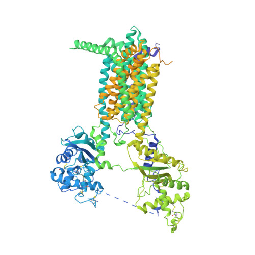
    The Dispatched protein, which is related to the NPC1 and PTCH1 cholesterol transporters 1,2 and to H + -driven transporters of the RND family 3,4 , enables tissue-patterning activity of the lipid-modified Hedgehog protein by releasing it from tightly -localized sites of embryonic expression 5-10 . Here we determine a cryo-electron microscopy structure of the mouse protein Dispatched homologue 1 (DISP1), revealing three Na + ions coordinated within a channel that traverses its transmembrane domain. We find that the rate of Hedgehog export is dependent on the Na + gradient across the plasma membrane. The transmembrane channel and Na + binding are disrupted in DISP1-NNN, a variant with asparagine substitutions for three intramembrane aspartate residues that each coordinate and neutralize the charge of one of the three Na + ions. DISP1-NNN and variants that disrupt single Na + sites retain binding to, but are impaired in export of the lipid-modified Hedgehog protein to the SCUBE2 acceptor. Interaction of the amino-terminal signalling domain of the Sonic hedgehog protein (ShhN) with DISP1 occurs via an extensive buried surface area and contacts with an extended furin-cleaved DISP1 arm. Variability analysis reveals that ShhN binding is restricted to one extreme of a continuous series of DISP1 conformations. The bound and unbound DISP1 conformations display distinct Na + -site occupancies, which suggests a mechanism by which transmembrane Na + flux may power extraction of the lipid-linked Hedgehog signal from the membrane. Na + -coordinating residues in DISP1 are conserved in PTCH1 and other metazoan RND family members, suggesting that Na + flux powers their conformationally driven activities.


  • Organizational Affiliation

    Institute for Stem Cell Biology and Regenerative Medicine, Stanford University School of Medicine, Stanford, CA, USA.


Macromolecules
Find similar proteins by:  (by identity cutoff)  |  3D Structure
Entity ID: 1
MoleculeChains Sequence LengthOrganismDetailsImage
Protein dispatched homolog 11,352Mus musculusMutation(s): 0 
Gene Names: Disp1DispDispaIcbIcbins
Membrane Entity: Yes 
UniProt
Find proteins for Q3TDN0 (Mus musculus)
Explore Q3TDN0 
Go to UniProtKB:  Q3TDN0
Entity Groups  
Sequence Clusters30% Identity50% Identity70% Identity90% Identity95% Identity100% Identity
UniProt GroupQ3TDN0
Sequence Annotations
Expand
  • Reference Sequence
Small Molecules
Ligands 4 Unique
IDChains Name / Formula / InChI Key2D Diagram3D Interactions
AV0 (Subject of Investigation/LOI)
Query on AV0

Download Ideal Coordinates CCD File 
BA [auth A],
CA [auth A]
Lauryl Maltose Neopentyl Glycol
C47 H88 O22
MADJBYLAYPCCOO-VWHTXWAPSA-N
Y01 (Subject of Investigation/LOI)
Query on Y01

Download Ideal Coordinates CCD File 
AA [auth A]
B [auth A]
C [auth A]
D [auth A]
E [auth A]
AA [auth A],
B [auth A],
C [auth A],
D [auth A],
E [auth A],
F [auth A],
G [auth A],
H [auth A],
I [auth A],
J [auth A],
K [auth A],
L [auth A],
M [auth A],
N [auth A],
O [auth A],
P [auth A],
Q [auth A],
R [auth A],
S [auth A],
T [auth A],
U [auth A],
V [auth A],
W [auth A],
X [auth A],
Y [auth A],
Z [auth A]
CHOLESTEROL HEMISUCCINATE
C31 H50 O4
WLNARFZDISHUGS-MIXBDBMTSA-N
NAG
Query on NAG

Download Ideal Coordinates CCD File 
DA [auth A],
EA [auth A],
FA [auth A],
GA [auth A],
HA [auth A]
2-acetamido-2-deoxy-beta-D-glucopyranose
C8 H15 N O6
OVRNDRQMDRJTHS-FMDGEEDCSA-N
NA (Subject of Investigation/LOI)
Query on NA

Download Ideal Coordinates CCD File 
IA [auth A],
JA [auth A]
SODIUM ION
Na
FKNQFGJONOIPTF-UHFFFAOYSA-N
Experimental Data & Validation

Experimental Data

  • Method: ELECTRON MICROSCOPY
  • Resolution: 2.50 Å
  • Aggregation State: PARTICLE 
  • Reconstruction Method: SINGLE PARTICLE 
EM Software:
TaskSoftware PackageVersion
RECONSTRUCTIONcryoSPARC
MODEL REFINEMENTPHENIX
MODEL REFINEMENTISOLDE

Structure Validation

View Full Validation Report


Entry History & Funding Information

Deposition Data


Funding OrganizationLocationGrant Number
National Institutes of Health/National Institute of General Medical Sciences (NIH/NIGMS)United StatesR01GM102498
National Institutes of Health/National Institute of General Medical Sciences (NIH/NIGMS)United StatesR35GM140847
National Institutes of Health/National Institute of General Medical Sciences (NIH/NIGMS)United StatesS10OD020054
National Institutes of Health/National Institute of General Medical Sciences (NIH/NIGMS)United StatesS10OD021741
Howard Hughes Medical Institute (HHMI)United States--

Revision History  (Full details and data files)

  • Version 1.0: 2021-10-27
    Type: Initial release
  • Version 1.1: 2021-11-10
    Changes: Database references
  • Version 1.2: 2021-11-24
    Changes: Database references
  • Version 1.3: 2023-12-13
    Changes: Data collection, Derived calculations, Structure summary