7S6H

Human PARP1 deltaV687-E688 bound to NAD+ analog EB-47 and to a DNA double strand break.


Experimental Data Snapshot

  • Method: X-RAY DIFFRACTION
  • Resolution: 3.10 Å
  • R-Value Free: 0.251 
  • R-Value Work: 0.210 
  • R-Value Observed: 0.212 

wwPDB Validation   3D Report Full Report


Ligand Structure Quality Assessment 


This is version 1.3 of the entry. See complete history


Literature

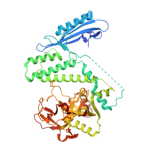
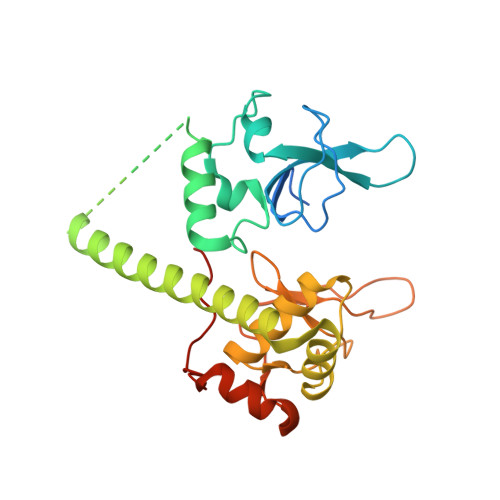
Captured snapshots of PARP1 in the active state reveal the mechanics of PARP1 allostery.

Rouleau-Turcotte, E.Krastev, D.B.Pettitt, S.J.Lord, C.J.Pascal, J.M.

(2022) Mol Cell 82: 2939-2951.e5

  • DOI: https://doi.org/10.1016/j.molcel.2022.06.011
  • Primary Citation of Related Structures:  
    7S68, 7S6H, 7S6M, 7S81

  • PubMed Abstract: 

    PARP1 rapidly detects DNA strand break damage and allosterically signals break detection to the PARP1 catalytic domain to activate poly(ADP-ribose) production from NAD + . PARP1 activation is characterized by dynamic changes in the structure of a regulatory helical domain (HD); yet, there are limited insights into the specific contributions that the HD makes to PARP1 allostery. Here, we have determined crystal structures of PARP1 in isolated active states that display specific HD conformations. These captured snapshots and biochemical analysis illustrate HD contributions to PARP1 multi-domain and high-affinity interaction with DNA damage, provide novel insights into the mechanics of PARP1 allostery, and indicate how HD active conformations correspond to alterations in the catalytic region that reveal the active site to NAD + . Our work deepens the understanding of PARP1 catalytic activation, the dynamics of the binding site of PARP inhibitor compounds, and the mechanisms regulating PARP1 retention on DNA damage.


  • Organizational Affiliation

    Department of Biochemistry and Molecular Medicine, Université de Montréal, Montréal, QC H3T 1J4, Canada.


Macromolecules

Find similar proteins by:  (by identity cutoff)  |  3D Structure
Entity ID: 3
MoleculeChains Sequence LengthOrganismDetailsImage
Poly [ADP-ribose] polymerase 1D [auth B],
H [auth D]
504Homo sapiensMutation(s): 1 
Gene Names: PARP1ADPRTPPOL
EC: 2.4.2.30 (PDB Primary Data), 2.4.2 (PDB Primary Data)
UniProt & NIH Common Fund Data Resources
Find proteins for P09874 (Homo sapiens)
Explore P09874 
Go to UniProtKB:  P09874
PHAROS:  P09874
GTEx:  ENSG00000143799 
Entity Groups  
Sequence Clusters30% Identity50% Identity70% Identity90% Identity95% Identity100% Identity
UniProt GroupP09874
Sequence Annotations
Expand
  • Reference Sequence
Find similar proteins by:  (by identity cutoff)  |  3D Structure
Entity ID: 4
MoleculeChains Sequence LengthOrganismDetailsImage
Fusion of human PARP1 zinc fingers 1 and 3 (Zn1, Zn3)F [auth A],
G [auth C]
276Homo sapiensMutation(s): 0 
Gene Names: PARP1ADPRTPPOL
EC: 2.4.2.30 (PDB Primary Data), 2.4.2 (PDB Primary Data)
UniProt & NIH Common Fund Data Resources
Find proteins for P09874 (Homo sapiens)
Explore P09874 
Go to UniProtKB:  P09874
PHAROS:  P09874
GTEx:  ENSG00000143799 
Entity Groups  
Sequence Clusters30% Identity50% Identity70% Identity90% Identity95% Identity100% Identity
UniProt GroupP09874
Sequence Annotations
Expand
  • Reference Sequence

Find similar nucleic acids by:  Sequence   |   3D Structure  

Entity ID: 1
MoleculeChains LengthOrganismImage
DNA (5'-D(*CP*GP*AP*CP*G)-3')A [auth M],
C [auth E]
5synthetic construct
Sequence Annotations
Expand
  • Reference Sequence

Find similar nucleic acids by:  Sequence   |   3D Structure  

Entity ID: 2
MoleculeChains LengthOrganismImage
DNA (5'-D(*CP*GP*TP*CP*G)-3')B [auth N],
E [auth F]
5synthetic construct
Sequence Annotations
Expand
  • Reference Sequence
Small Molecules
Ligands 3 Unique
IDChains Name / Formula / InChI Key2D Diagram3D Interactions
UHB (Subject of Investigation/LOI)
Query on UHB

Download Ideal Coordinates CCD File 
J [auth B],
P [auth D]
2-[4-[(2S,3S,4R,5R)-5-(6-aminopurin-9-yl)-3,4-bis(oxidanyl)oxolan-2-yl]carbonylpiperazin-1-yl]-N-(1-oxidanylidene-2,3-dihydroisoindol-4-yl)ethanamide
C24 H27 N9 O6
QEUOCRGVJJDDTK-VBFAUCSFSA-N
ZN
Query on ZN

Download Ideal Coordinates CCD File 
K [auth A],
L [auth A],
M [auth C],
N [auth C]
ZINC ION
Zn
PTFCDOFLOPIGGS-UHFFFAOYSA-N
EDO
Query on EDO

Download Ideal Coordinates CCD File 
I [auth B],
O [auth D]
1,2-ETHANEDIOL
C2 H6 O2
LYCAIKOWRPUZTN-UHFFFAOYSA-N
Experimental Data & Validation

Experimental Data

  • Method: X-RAY DIFFRACTION
  • Resolution: 3.10 Å
  • R-Value Free: 0.251 
  • R-Value Work: 0.210 
  • R-Value Observed: 0.212 
  • Space Group: P 1 21 1
Unit Cell:
Length ( Å )Angle ( ˚ )
a = 111.467α = 90
b = 110.651β = 115.06
c = 116.475γ = 90
Software Package:
Software NamePurpose
PHENIXrefinement
Aimlessdata scaling
PDB_EXTRACTdata extraction
XDSdata reduction
PHASERphasing

Structure Validation

View Full Validation Report


Ligand Structure Quality Assessment 


Entry History & Funding Information

Deposition Data


Funding OrganizationLocationGrant Number
Canadian Institutes of Health Research (CIHR)CanadaPJT173370

Revision History  (Full details and data files)

  • Version 1.0: 2022-06-29
    Type: Initial release
  • Version 1.1: 2022-07-20
    Changes: Database references
  • Version 1.2: 2022-08-31
    Changes: Database references
  • Version 1.3: 2023-10-18
    Changes: Data collection, Refinement description