7TIM | pdb_00007tim

STRUCTURE OF THE TRIOSEPHOSPHATE ISOMERASE-PHOSPHOGLYCOLOHYDROXAMATE COMPLEX: AN ANALOGUE OF THE INTERMEDIATE ON THE REACTION PATHWAY


Experimental Data Snapshot

  • Method: X-RAY DIFFRACTION
  • Resolution: 1.90 Å
  • R-Value Work: 
    0.183 (Depositor) 
  • R-Value Observed: 
    0.183 (Depositor) 

wwPDB Validation 3D Report Full Report

Validation slider image for 7TIM

This is version 1.3 of the entry. See complete history

Literature

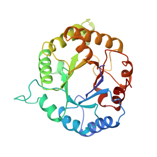
Structure of the triosephosphate isomerase-phosphoglycolohydroxamate complex: an analogue of the intermediate on the reaction pathway.

Davenport, R.C.Bash, P.A.Seaton, B.A.Karplus, M.Petsko, G.A.Ringe, D.

(1991) Biochemistry 30: 5821-5826

  • DOI: https://doi.org/10.1021/bi00238a002
  • Primary Citation Related Structures: 
    7TIM

  • PubMed Abstract: 

    The glycolytic enzyme triosephosphate isomerase (TIM) catalyzes the interconversion of the three-carbon sugars dihydroxyacetone phosphate (DHAP) and D-glyceraldehyde 3-phosphate (GAP) at a rate limited by the diffusion of substrate to the enzyme. We have solved the three-dimensional structure of TIM complexed with a reactive intermediate analogue, phosphoglycolohydroxamate (PGH), at 1.9-A resolution and have refined the structure to an R-factor of 18%. Analysis of the refined structure reveals the geometry of the active-site residues and the interactions they make with the inhibitor and, by analogy, the substrates. The structure is consistent with an acid-base mechanism in which the carboxylate of Glu-165 abstracts a proton from carbon while His-95 donates a proton to oxygen to form an enediol (or enediolate) intermediate. The conformation of the bound substrate stereoelectronically favors proton transfer from substrate carbon to the syn orbital of Glu-165. The crystal structure suggests that His-95 is neutral rather than cationic in the ground state and therefore would have to function as an imidazole acid instead of the usual imidazolium. Lys-12 is oriented so as to polarize the substrate oxygens by hydrogen bonding and/or electrostatic interaction, providing stabilization for the charged transition state. Asn-10 may play a similar role.


  • Organizational Affiliation
    • Department of Chemistry, Massachusetts Institute of Technology, Cambridge 02139.

Macromolecule Content 

  • Total Structure Weight: 53.73 kDa 
  • Atom Count: 4,033 
  • Modeled Residue Count: 494 
  • Deposited Residue Count: 494 
  • Unique protein chains: 1

Macromolecules

Find similar proteins by:|  3D Structure
Entity ID: 1
MoleculeChains  Sequence LengthOrganismDetailsImage
TRIOSEPHOSPHATE ISOMERASE
A, B
247Saccharomyces cerevisiaeMutation(s): 0 
EC: 5.3.1.1
UniProt
Find proteins for P00942 (Saccharomyces cerevisiae (strain ATCC 204508 / S288c))
Explore P00942 
Go to UniProtKB:  P00942
Entity Groups
Sequence Clusters30% Identity50% Identity70% Identity90% Identity95% Identity100% Identity
UniProt GroupP00942
Sequence Annotations
Expand
Reference Sequence

Small Molecules

Ligands 1 Unique
IDChains Name / Formula / InChI Key2D Diagram3D Interactions
PGH

Query on PGH


Download:Ideal Coordinates CCD File
C [auth A],
D [auth B]
PHOSPHOGLYCOLOHYDROXAMIC ACID
C2 H6 N O6 P
BAXHHWZKQZIJID-UHFFFAOYSA-N

Experimental Data & Validation

Experimental Data

  • Method: X-RAY DIFFRACTION
  • Resolution: 1.90 Å
  • R-Value Work:  0.183 (Depositor) 
  • R-Value Observed: 0.183 (Depositor) 
Space Group: P 1 21 1
Unit Cell:
Length ( Å )Angle ( ˚ )
a = 74α = 90
b = 83.5β = 99.55
c = 38.4γ = 90
Software Package:
Software NamePurpose
X-PLORmodel building
PROLSQrefinement
X-PLORrefinement
X-PLORphasing

Structure Validation

View Full Validation Report


Entry History 

Deposition Data

Revision History  (Full details and data files)

  • Version 1.0: 1993-10-31
    Type: Initial release
  • Version 1.1: 2008-03-25
    Changes: Version format compliance
  • Version 1.2: 2011-07-13
    Changes: Version format compliance
  • Version 1.3: 2024-02-28
    Changes: Data collection, Database references, Derived calculations, Other