7TO5 | pdb_00007to5

HIV-1 wild type protease with GRL-05816A, with C-4 substituted cyclohexane-fused bis-tetrahydrofuran (Chf-THF) derivatives as P2-ligand [diastereomer 1]


Experimental Data Snapshot

  • Method: X-RAY DIFFRACTION
  • Resolution: 1.13 Å
  • R-Value Free: 
    0.142 (Depositor), 0.143 (DCC) 
  • R-Value Work: 
    0.123 (Depositor), 0.124 (DCC) 
  • R-Value Observed: 
    0.124 (Depositor) 

Starting Model: experimental
View more details

wwPDB Validation 3D Report Full Report

Validation slider image for 7TO5

Ligand Structure Quality Assessment 


This is version 1.3 of the entry. See complete history

Literature

Design, Synthesis and X-Ray Structural Studies of Potent HIV-1 Protease Inhibitors Containing C-4 Substituted Tricyclic Hexahydro-Furofuran Derivatives as P2 Ligands.

Ghosh, A.K.Kovela, S.Sharma, A.Shahabi, D.Ghosh, A.K.Hopkins, D.R.Yadav, M.Johnson, M.E.Agniswamy, J.Wang, Y.F.Hattori, S.I.Higashi-Kuwata, N.Aoki, M.Amano, M.Weber, I.T.Mitsuya, H.

(2022) ChemMedChem 17: e202200058-e202200058

  • DOI: https://doi.org/10.1002/cmdc.202200058
  • Primary Citation Related Structures: 
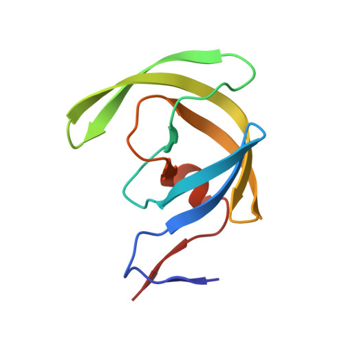
    7TO5, 7TO6

  • PubMed Abstract: 

    The design, synthesis, X-ray structural, and biological evaluation of a series of highly potent HIV-1 protease inhibitors are reported herein. These inhibitors incorporate novel cyclohexane-fused tricyclic bis-tetrahydrofuran as P2 ligands in combination with a variety of P1 and P2' ligands. The inhibitor with a difluoromethylphenyl P1 ligand and a cyclopropylaminobenzothiazole P2' ligand exhibited the most potent antiviral activity. Also, it maintained potent antiviral activity against a panel of highly multidrug-resistant HIV-1 variants. The corresponding inhibitor with an enantiomeric ligand was significantly less potent in these antiviral assays. The new P2 ligands were synthesized in optically active form using enzymatic desymmetrization of meso-diols as the key step. To obtain molecular insight, two high-resolution X-ray structures of inhibitor-bound HIV-1 protease were determined and structural analyses have been highlighted.


  • Organizational Affiliation
    • Department of Chemistry and Department of Medicinal Chemistry, Purdue University, West Lafayette, IN 47907, USA.

Macromolecule Content 

  • Total Structure Weight: 22.42 kDa 
  • Atom Count: 2,223 
  • Modeled Residue Count: 198 
  • Deposited Residue Count: 198 
  • Unique protein chains: 1

Macromolecules

Find similar proteins by:|  3D Structure
Entity ID: 1
MoleculeChains  Sequence LengthOrganismDetailsImage
Protease
A, B
99Human immunodeficiency virus 1Mutation(s): 5 
Gene Names: pol
UniProt
Find proteins for P03367 (Human immunodeficiency virus type 1 group M subtype B (isolate BRU/LAI))
Explore P03367 
Go to UniProtKB:  P03367
Entity Groups
Sequence Clusters30% Identity50% Identity70% Identity90% Identity95% Identity100% Identity
UniProt GroupP03367
Sequence Annotations
Expand
Reference Sequence

Small Molecules

Ligands 5 Unique
IDChains Name / Formula / InChI Key2D Diagram3D Interactions
G8R
(Subject of Investigation/LOI)

Query on G8R


Download:Ideal Coordinates CCD File
F [auth B](1R,3aS,4S,6S,7aR)-octahydro-1,6-epoxy-2-benzofuran-4-yl [(2S,3R)-4-{[2-(cyclopropylamino)-1,3-benzothiazole-6-sulfonyl](2-methylpropyl)amino}-3-hydroxy-1-phenylbutan-2-yl]carbamate
C33 H42 N4 O7 S2
QVXBJENHAYPWAW-XGNLSKQLSA-N
GOL

Query on GOL


Download:Ideal Coordinates CCD File
I [auth B]GLYCEROL
C3 H8 O3
PEDCQBHIVMGVHV-UHFFFAOYSA-N
FMT

Query on FMT


Download:Ideal Coordinates CCD File
E [auth A]FORMIC ACID
C H2 O2
BDAGIHXWWSANSR-UHFFFAOYSA-N
CL

Query on CL


Download:Ideal Coordinates CCD File
D [auth A],
G [auth B],
H [auth B]
CHLORIDE ION
Cl
VEXZGXHMUGYJMC-UHFFFAOYSA-M
NA

Query on NA


Download:Ideal Coordinates CCD File
C [auth A]SODIUM ION
Na
FKNQFGJONOIPTF-UHFFFAOYSA-N

Experimental Data & Validation

Experimental Data

  • Method: X-RAY DIFFRACTION
  • Resolution: 1.13 Å
  • R-Value Free:  0.142 (Depositor), 0.143 (DCC) 
  • R-Value Work:  0.123 (Depositor), 0.124 (DCC) 
  • R-Value Observed: 0.124 (Depositor) 
Space Group: P 21 21 2
Unit Cell:
Length ( Å )Angle ( ˚ )
a = 58.943α = 90
b = 86.138β = 90
c = 46.097γ = 90
Software Package:
Software NamePurpose
HKL-2000data reduction
HKL-2000data scaling
PHASERphasing
REFMACrefinement
PDB_EXTRACTdata extraction

Structure Validation

View Full Validation Report


Ligand Structure Quality Assessment 


Entry History 

& Funding Information

Deposition Data


Funding OrganizationLocationGrant Number
National Institutes of Health/National Institute Of Allergy and Infectious Diseases (NIH/NIAID)United StatesAI150466
National Institutes of Health/National Institute Of Allergy and Infectious Diseases (NIH/NIAID)United StatesAI150461

Revision History  (Full details and data files)

  • Version 1.0: 2022-03-02
    Type: Initial release
  • Version 1.1: 2022-03-16
    Changes: Database references
  • Version 1.2: 2022-05-18
    Changes: Database references
  • Version 1.3: 2023-10-18
    Changes: Data collection, Refinement description