7UBY

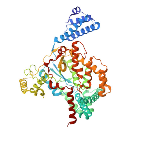
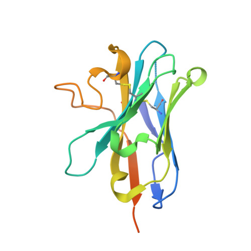
Structure of the GTD domain of Clostridium difficile toxin A in complex with VHH AH3


Experimental Data Snapshot

  • Method: X-RAY DIFFRACTION
  • Resolution: 2.10 Å
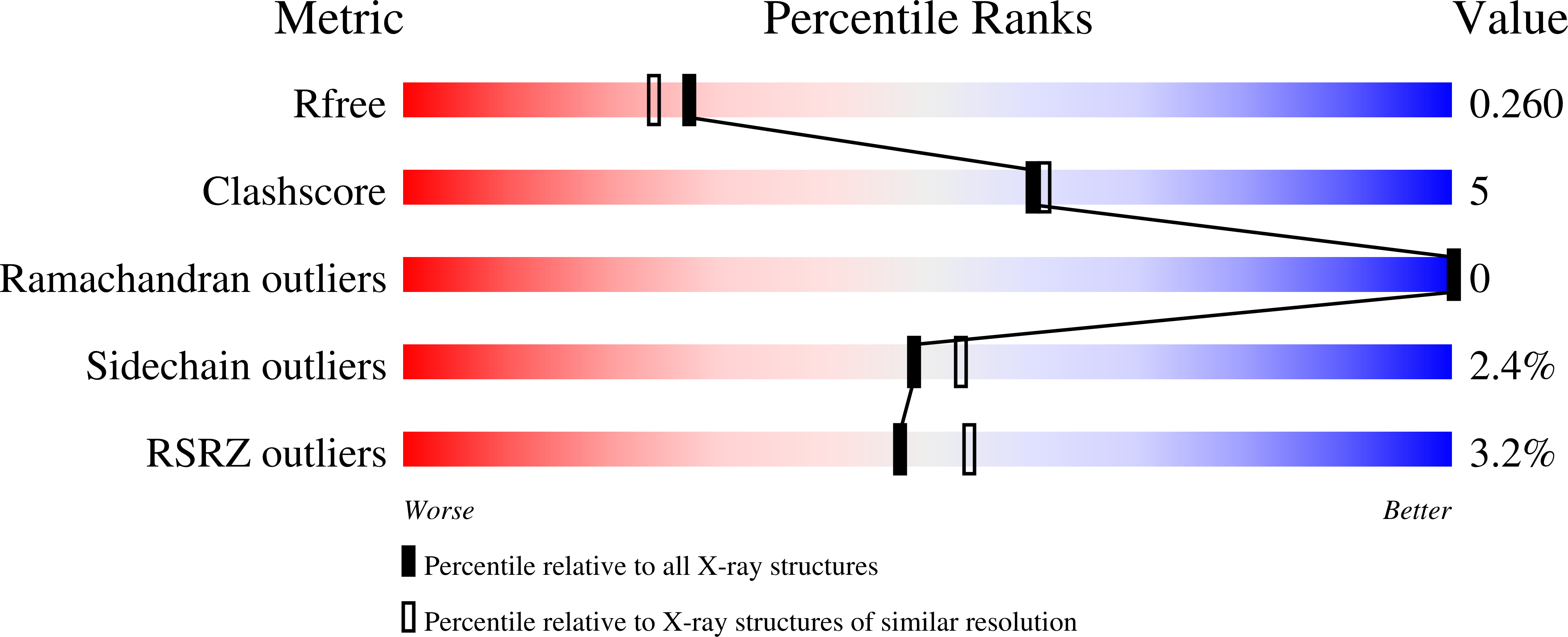
  • R-Value Free: 0.261 
  • R-Value Work: 0.218 
  • R-Value Observed: 0.219 

Starting Model: experimental
View more details

wwPDB Validation   3D Report Full Report


Ligand Structure Quality Assessment 


This is version 1.2 of the entry. See complete history


Literature

Neutralizing epitopes on Clostridioides difficile toxin A revealed by the structures of two camelid VHH antibodies.

Chen, B.Perry, K.Jin, R.

(2022) Front Immunol 13: 978858-978858

  • DOI: https://doi.org/10.3389/fimmu.2022.978858
  • Primary Citation of Related Structures:  
    7UBX, 7UBY

  • PubMed Abstract: 

    Toxin A (TcdA) and toxin B (TcdB) are two key virulence factors secreted by Clostridioides difficile , which is listed as an urgent threat by the CDC. These two large homologous exotoxins are mainly responsible for diseases associated with C. difficile infection (CDI) with symptoms ranging from diarrhea to life threatening pseudomembranous colitis. Single-domain camelid antibodies (VHHs) AH3 and AA6 are two potent antitoxins against TcdA, which when combined with two TcdB-targeting VHHs showed effective protection against both primary and recurrent CDI in animal models. Here, we report the co-crystal structures of AH3 and AA6 when they form complexes with the glucosyltransferase domain (GTD) and a fragment of the delivery and receptor-binding domain (DRBD) of TcdA, respectively. Based on these structures, we find that AH3 binding enhances the overall stability of the GTD and interferes with its unfolding at acidic pH, and AA6 may inhibit the pH-dependent conformational changes in the DRBD that is necessary for pore formation of TcdA. These studies reveal two functionally critical epitopes on TcdA and shed new insights into neutralizing mechanisms and potential development of epitope-focused vaccines against TcdA.


  • Organizational Affiliation

    Department of Physiology and Biophysics, School of Medicine, University of California, Irvine, CA, United States.


Macromolecules
Find similar proteins by:  (by identity cutoff)  |  3D Structure
Entity ID: 1
MoleculeChains Sequence LengthOrganismDetailsImage
Glucosyltransferase TcdAA [auth B],
C [auth A]
543Clostridioides difficileMutation(s): 1 
Gene Names: tcdAtoxA
EC: 2.4.1 (PDB Primary Data), 3.4.22 (UniProt)
UniProt
Find proteins for P16154 (Clostridioides difficile)
Explore P16154 
Go to UniProtKB:  P16154
Entity Groups  
Sequence Clusters30% Identity50% Identity70% Identity90% Identity95% Identity100% Identity
UniProt GroupP16154
Sequence Annotations
Expand
  • Reference Sequence
Find similar proteins by:  (by identity cutoff)  |  3D Structure
Entity ID: 2
MoleculeChains Sequence LengthOrganismDetailsImage
Nanobody VHH AH3B [auth D],
D [auth C]
151CamelidaeMutation(s): 0 
Entity Groups  
Sequence Clusters30% Identity50% Identity70% Identity90% Identity95% Identity100% Identity
Sequence Annotations
Expand
  • Reference Sequence
Small Molecules
Ligands 4 Unique
IDChains Name / Formula / InChI Key2D Diagram3D Interactions
UPG (Subject of Investigation/LOI)
Query on UPG

Download Ideal Coordinates CCD File 
E [auth B],
M [auth A]
URIDINE-5'-DIPHOSPHATE-GLUCOSE
C15 H24 N2 O17 P2
HSCJRCZFDFQWRP-JZMIEXBBSA-N
PEG
Query on PEG

Download Ideal Coordinates CCD File 
G [auth B],
H [auth B]
DI(HYDROXYETHYL)ETHER
C4 H10 O3
MTHSVFCYNBDYFN-UHFFFAOYSA-N
EDO
Query on EDO

Download Ideal Coordinates CCD File 
I [auth B]
J [auth B]
K [auth B]
L [auth B]
O [auth A]
I [auth B],
J [auth B],
K [auth B],
L [auth B],
O [auth A],
P [auth A],
Q [auth A],
R [auth A],
S [auth A],
T [auth A],
U [auth A]
1,2-ETHANEDIOL
C2 H6 O2
LYCAIKOWRPUZTN-UHFFFAOYSA-N
MN
Query on MN

Download Ideal Coordinates CCD File 
F [auth B],
N [auth A]
MANGANESE (II) ION
Mn
WAEMQWOKJMHJLA-UHFFFAOYSA-N
Experimental Data & Validation

Experimental Data

  • Method: X-RAY DIFFRACTION
  • Resolution: 2.10 Å
  • R-Value Free: 0.261 
  • R-Value Work: 0.218 
  • R-Value Observed: 0.219 
  • Space Group: P 1 21 1
Unit Cell:
Length ( Å )Angle ( ˚ )
a = 80.118α = 90
b = 131.641β = 110.015
c = 83.796γ = 90
Software Package:
Software NamePurpose
PHENIXrefinement
XDSdata scaling
PHENIXphasing

Structure Validation

View Full Validation Report


Ligand Structure Quality Assessment 


Entry History & Funding Information

Deposition Data


Funding OrganizationLocationGrant Number
National Institutes of Health/National Institute Of Allergy and Infectious Diseases (NIH/NIAID)United States--

Revision History  (Full details and data files)

  • Version 1.0: 2022-11-16
    Type: Initial release
  • Version 1.1: 2022-12-14
    Changes: Database references, Structure summary
  • Version 1.2: 2023-10-25
    Changes: Data collection, Refinement description