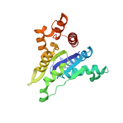
7UXS

Crystal structure of the BcThsA SLOG domain in complex with 3'cADPR


Experimental Data Snapshot

  • Method: X-RAY DIFFRACTION
  • Resolution: 1.57 Å
  • R-Value Free: 0.176 
  • R-Value Work: 0.149 
  • R-Value Observed: 0.150 

Starting Model: experimental
View more details

wwPDB Validation   3D Report Full Report


Ligand Structure Quality Assessment 


This is version 1.3 of the entry. See complete history


Literature

Cyclic ADP ribose isomers: Production, chemical structures, and immune signaling.

Manik, M.K.Shi, Y.Li, S.Zaydman, M.A.Damaraju, N.Eastman, S.Smith, T.G.Gu, W.Masic, V.Mosaiab, T.Weagley, J.S.Hancock, S.J.Vasquez, E.Hartley-Tassell, L.Kargios, N.Maruta, N.Lim, B.Y.J.Burdett, H.Landsberg, M.J.Schembri, M.A.Prokes, I.Song, L.Grant, M.DiAntonio, A.Nanson, J.D.Guo, M.Milbrandt, J.Ve, T.Kobe, B.

(2022) Science 377: eadc8969-eadc8969

  • DOI: https://doi.org/10.1126/science.adc8969
  • Primary Citation of Related Structures:  
    7UWG, 7UXR, 7UXS, 7UXT, 7UXU

  • PubMed Abstract: 

    Cyclic adenosine diphosphate (ADP)-ribose (cADPR) isomers are signaling molecules produced by bacterial and plant Toll/interleukin-1 receptor (TIR) domains via nicotinamide adenine dinucleotide (oxidized form) (NAD + ) hydrolysis. We show that v-cADPR (2'cADPR) and v2-cADPR (3'cADPR) isomers are cyclized by O-glycosidic bond formation between the ribose moieties in ADPR. Structures of 2'cADPR-producing TIR domains reveal conformational changes that lead to an active assembly that resembles those of Toll-like receptor adaptor TIR domains. Mutagenesis reveals a conserved tryptophan that is essential for cyclization. We show that 3'cADPR is an activator of ThsA effector proteins from the bacterial antiphage defense system termed Thoeris and a suppressor of plant immunity when produced by the effector HopAM1. Collectively, our results reveal the molecular basis of cADPR isomer production and establish 3'cADPR in bacteria as an antiviral and plant immunity-suppressing signaling molecule.


  • Organizational Affiliation

    School of Chemistry and Molecular Biosciences, The University of Queensland, Brisbane, QLD 4072, Australia.


Macromolecules
Find similar proteins by:  (by identity cutoff)  |  3D Structure
Entity ID: 1
MoleculeChains Sequence LengthOrganismDetailsImage
BcThsAA [auth B],
B [auth A]
196Bacillus subtilisMutation(s): 0 
EC: 3.2.2.5
UniProt
Find proteins for J8G6Z1 (Bacillus cereus (strain MSX-D12))
Explore J8G6Z1 
Go to UniProtKB:  J8G6Z1
Entity Groups  
Sequence Clusters30% Identity50% Identity70% Identity90% Identity95% Identity100% Identity
UniProt GroupJ8G6Z1
Sequence Annotations
Expand
  • Reference Sequence
Small Molecules
Ligands 3 Unique
IDChains Name / Formula / InChI Key2D Diagram3D Interactions
OJC (Subject of Investigation/LOI)
Query on OJC

Download Ideal Coordinates CCD File 
C [auth B],
F [auth A]
(2R,3R,3aS,5S,6R,7S,8R,11R,13S,15aR)-2-(6-amino-9H-purin-9-yl)-3,6,7,11,13-pentahydroxyoctahydro-2H,5H,11H,13H-5,8-epoxy-11lambda~5~,13lambda~5~-furo[2,3-g][1,3,5,9,2,4]tetraoxadiphosphacyclotetradecine-11,13-dione
C15 H21 N5 O13 P2
NDSLKXAJVDJCEG-ZQSHOCFMSA-N
SO4
Query on SO4

Download Ideal Coordinates CCD File 
E [auth B]SULFATE ION
O4 S
QAOWNCQODCNURD-UHFFFAOYSA-L
GOL
Query on GOL

Download Ideal Coordinates CCD File 
D [auth B],
G [auth A]
GLYCEROL
C3 H8 O3
PEDCQBHIVMGVHV-UHFFFAOYSA-N
Experimental Data & Validation

Experimental Data

  • Method: X-RAY DIFFRACTION
  • Resolution: 1.57 Å
  • R-Value Free: 0.176 
  • R-Value Work: 0.149 
  • R-Value Observed: 0.150 
  • Space Group: P 21 21 21
Unit Cell:
Length ( Å )Angle ( ˚ )
a = 46.194α = 90
b = 70.801β = 90
c = 121.74γ = 90
Software Package:
Software NamePurpose
PHENIXrefinement
XDSdata reduction
Aimlessdata scaling
PHASERphasing

Structure Validation

View Full Validation Report


Ligand Structure Quality Assessment 


Entry History & Funding Information

Deposition Data


Funding OrganizationLocationGrant Number
Australian Research Council (ARC)Australia--
National Health and Medical Research Council (NHMRC, Australia)Australia--

Revision History  (Full details and data files)

  • Version 1.0: 2022-09-07
    Type: Initial release
  • Version 1.1: 2022-09-14
    Changes: Database references
  • Version 1.2: 2022-10-12
    Changes: Database references
  • Version 1.3: 2023-10-18
    Changes: Data collection, Refinement description