7Y6D | pdb_00007y6d

Cryo-EM structure of SARS-CoV-2 Delta variant spike proteins on intact virions: 3 Closed RBD


Experimental Data Snapshot

  • Method: ELECTRON MICROSCOPY
  • Resolution: 4.39 Å
  • Aggregation State: PARTICLE 
  • Reconstruction Method: SINGLE PARTICLE 

Starting Models: experimental
View more details

wwPDB Validation 3D Report Full Report

Validation slider image for 7Y6D

This is version 1.1 of the entry. See complete history

Literature

In situ architecture and membrane fusion of SARS-CoV-2 Delta variant.

Song, Y.Yao, H.Wu, N.Xu, J.Zhang, Z.Peng, C.Li, S.Kong, W.Chen, Y.Zhu, M.Wang, J.Shi, D.Zhao, C.Lu, X.Echavarria Galindo, M.Li, S.

(2023) Proc Natl Acad Sci U S A 120: e2213332120-e2213332120

  • DOI: https://doi.org/10.1073/pnas.2213332120
  • Primary Citation Related Structures: 
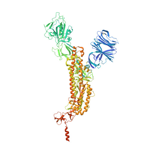
    7Y6D

  • PubMed Abstract: 

    Among the current five Variants of Concern, infections caused by SARS-CoV-2 B.1.617.2 (Delta) variant are often associated with the greatest severity. Despite recent advances on the molecular basis of elevated pathogenicity using recombinant proteins, the architecture of intact Delta virions remains veiled. Moreover, pieces of molecular evidence for the detailed mechanism of S-mediated membrane fusion are missing. Here, we showed the pleomorphic nature of Delta virions from electron beam inactivated samples and reported the in situ structure and distribution of S on the authentic Delta variant. We also captured the virus-virus fusion events, which provided pieces of structural evidence for Delta's attenuated dependency on cellular factors for fusion activation, and proposed a model of S-mediated membrane fusion. Besides, site-specific glycan analysis revealed increased oligomannose-type glycosylation of native Delta S than that of the WT S. Together, these results disclose distinctive factors of Delta being the most virulent SARS-CoV-2 variant.


  • Organizational Affiliation
    • State Key Laboratory of Membrane Biology & Beijing Frontier Research Center for Biological Structure, School of Life Sciences, Tsinghua University, Beijing 100084, China.

Macromolecule Content 

  • Total Structure Weight: 438.12 kDa 
  • Atom Count: 24,651 
  • Modeled Residue Count: 3,036 
  • Deposited Residue Count: 3,819 
  • Unique protein chains: 1

Macromolecules

Find similar proteins by:|  3D Structure
Entity ID: 1
MoleculeChains  Sequence LengthOrganismDetailsImage
Spike glycoprotein
A, B, C
1,273Severe acute respiratory syndrome coronavirus 2Mutation(s): 7 
UniProt
Find proteins for P0DTC2 (Severe acute respiratory syndrome coronavirus 2)
Explore P0DTC2 
Go to UniProtKB:  P0DTC2
Entity Groups
Sequence Clusters30% Identity50% Identity70% Identity90% Identity95% Identity100% Identity
UniProt GroupP0DTC2
Glycosylation
Glycosylation Sites: 16Go to GlyGen: P0DTC2-1
Sequence Annotations
Expand
Reference Sequence

Small Molecules

Ligands 1 Unique
IDChains Name / Formula / InChI Key2D Diagram3D Interactions
NAG

Query on NAG


Download:Ideal Coordinates CCD File
AA [auth A]
BA [auth A]
CA [auth A]
DA [auth B]
EA [auth B]
AA [auth A],
BA [auth A],
CA [auth A],
DA [auth B],
EA [auth B],
FA [auth B],
GA [auth B],
HA [auth B],
IA [auth B],
JA [auth B],
KA [auth B],
LA [auth B],
MA [auth B],
NA [auth B],
OA [auth C],
PA [auth C],
QA [auth C],
RA [auth C],
S [auth A],
SA [auth C],
T [auth A],
TA [auth C],
U [auth A],
UA [auth C],
V [auth A],
VA [auth C],
W [auth A],
WA [auth C],
X [auth A],
XA [auth C],
Y [auth A],
YA [auth C],
Z [auth A]
2-acetamido-2-deoxy-beta-D-glucopyranose
C8 H15 N O6
OVRNDRQMDRJTHS-FMDGEEDCSA-N

Experimental Data & Validation

Experimental Data

  • Method: ELECTRON MICROSCOPY
  • Resolution: 4.39 Å
  • Aggregation State: PARTICLE 
  • Reconstruction Method: SINGLE PARTICLE 
EM Software:
TaskSoftware PackageVersion
MODEL REFINEMENTPHENIX1.20.1
RECONSTRUCTIONRELION3.1.0

Structure Validation

View Full Validation Report


Entry History 

& Funding Information

Deposition Data


Funding OrganizationLocationGrant Number
National Natural Science Foundation of China (NSFC)China32171195

Revision History  (Full details and data files)

  • Version 1.0: 2023-06-07
    Type: Initial release
  • Version 1.1: 2024-11-06
    Changes: Data collection, Refinement description, Structure summary