7ZLS

co-crystal structure of SOCS2:ElonginB:ElonginC in complex with compound 13


Experimental Data Snapshot

  • Method: X-RAY DIFFRACTION
  • Resolution: 1.92 Å
  • R-Value Free: 0.238 
  • R-Value Work: 0.202 
  • R-Value Observed: 0.204 

Starting Model: other
View more details

wwPDB Validation   3D Report Full Report


Ligand Structure Quality Assessment 


This is version 1.3 of the entry. See complete history


Literature

Structure-based design of a phosphotyrosine-masked covalent ligand targeting the E3 ligase SOCS2.

Ramachandran, S.Makukhin, N.Haubrich, K.Nagala, M.Forrester, B.Lynch, D.M.Casement, R.Testa, A.Bruno, E.Gitto, R.Ciulli, A.

(2023) Nat Commun 14: 6345-6345

  • DOI: https://doi.org/10.1038/s41467-023-41894-3
  • Primary Citation of Related Structures:  
    7ZLM, 7ZLN, 7ZLO, 7ZLP, 7ZLR, 7ZLS

  • PubMed Abstract: 

    The Src homology 2 (SH2) domain recognizes phosphotyrosine (pY) post translational modifications in partner proteins to trigger downstream signaling. Drug discovery efforts targeting the SH2 domains have long been stymied by the poor drug-like properties of phosphate and its mimetics. Here, we use structure-based design to target the SH2 domain of the E3 ligase suppressor of cytokine signaling 2 (SOCS2). Starting from the highly ligand-efficient pY amino acid, a fragment growing approach reveals covalent modification of Cys111 in a co-crystal structure, which we leverage to rationally design a cysteine-directed electrophilic covalent inhibitor MN551. We report the prodrug MN714 containing a pivaloyloxymethyl (POM) protecting group and evidence its cell permeability and capping group unmasking using cellular target engagement and in-cell 19 F NMR spectroscopy. Covalent engagement at Cys111 competitively blocks recruitment of cellular SOCS2 protein to its native substrate. The qualified inhibitors of SOCS2 could find attractive applications as chemical probes to understand the biology of SOCS2 and its CRL5 complex, and as E3 ligase handles in proteolysis targeting chimera (PROTACs) to induce targeted protein degradation.


  • Organizational Affiliation

    Centre for Targeted Protein Degradation, Division of Biological Chemistry and Drug Discovery, School of Life Sciences, University of Dundee, 1 James Lindsay Place, Dundee, DD1 5JJ, United Kingdom.


Macromolecules
Find similar proteins by:  (by identity cutoff)  |  3D Structure
Entity ID: 1
MoleculeChains Sequence LengthOrganismDetailsImage
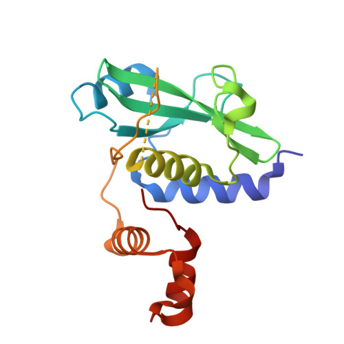
Suppressor of cytokine signaling 2A,
D,
G [auth J],
J [auth G]
169Homo sapiensMutation(s): 0 
Gene Names: SOCS2CIS2SSI2STATI2
UniProt & NIH Common Fund Data Resources
Find proteins for O14508 (Homo sapiens)
Explore O14508 
Go to UniProtKB:  O14508
PHAROS:  O14508
GTEx:  ENSG00000120833 
Entity Groups  
Sequence Clusters30% Identity50% Identity70% Identity90% Identity95% Identity100% Identity
UniProt GroupO14508
Sequence Annotations
Expand
  • Reference Sequence
Find similar proteins by:  (by identity cutoff)  |  3D Structure
Entity ID: 2
MoleculeChains Sequence LengthOrganismDetailsImage
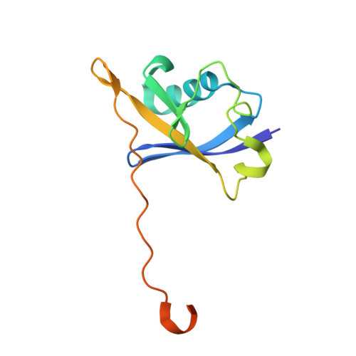
Elongin-BB,
E,
H [auth K],
K [auth H]
118Homo sapiensMutation(s): 0 
Gene Names: ELOBTCEB2
UniProt & NIH Common Fund Data Resources
Find proteins for Q15370 (Homo sapiens)
Explore Q15370 
Go to UniProtKB:  Q15370
PHAROS:  Q15370
GTEx:  ENSG00000103363 
Entity Groups  
Sequence Clusters30% Identity50% Identity70% Identity90% Identity95% Identity100% Identity
UniProt GroupQ15370
Sequence Annotations
Expand
  • Reference Sequence
Find similar proteins by:  (by identity cutoff)  |  3D Structure
Entity ID: 3
MoleculeChains Sequence LengthOrganismDetailsImage
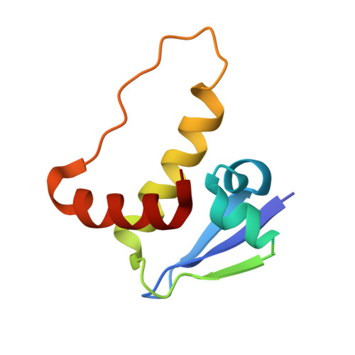
Elongin-CC,
F,
I [auth L],
L [auth I]
97Homo sapiensMutation(s): 0 
Gene Names: ELOCTCEB1
UniProt & NIH Common Fund Data Resources
Find proteins for Q15369 (Homo sapiens)
Explore Q15369 
Go to UniProtKB:  Q15369
PHAROS:  Q15369
GTEx:  ENSG00000154582 
Entity Groups  
Sequence Clusters30% Identity50% Identity70% Identity90% Identity95% Identity100% Identity
UniProt GroupQ15369
Sequence Annotations
Expand
  • Reference Sequence
Experimental Data & Validation

Experimental Data

  • Method: X-RAY DIFFRACTION
  • Resolution: 1.92 Å
  • R-Value Free: 0.238 
  • R-Value Work: 0.202 
  • R-Value Observed: 0.204 
  • Space Group: P 21 21 21
Unit Cell:
Length ( Å )Angle ( ˚ )
a = 59.549α = 90
b = 172.641β = 90
c = 185.475γ = 90
Software Package:
Software NamePurpose
Cootmodel building
PHENIXrefinement
xia2data reduction
xia2data scaling
PHASERphasing

Structure Validation

View Full Validation Report


Ligand Structure Quality Assessment 


Entry History & Funding Information

Deposition Data


Funding OrganizationLocationGrant Number
European Research Council (ERC)European UnionERC-2012-StG-311460 DrugE3CRLs
Innovative Medicines InitiativeSwitzerland875510

Revision History  (Full details and data files)

  • Version 1.0: 2023-04-26
    Type: Initial release
  • Version 1.1: 2023-10-11
    Changes: Data collection, Database references
  • Version 1.2: 2023-10-25
    Changes: Database references
  • Version 1.3: 2024-05-01
    Changes: Refinement description