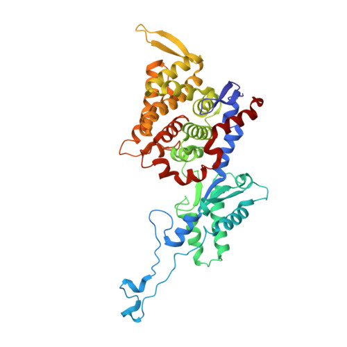
7B2C | pdb_00007b2c

Crystal structure of the ethyl-coenzyme M reductase from Candidatus Ethanoperedens thermophilum gassed with xenon


Experimental Data Snapshot

  • Method: X-RAY DIFFRACTION
  • Resolution: 1.80 Å
  • R-Value Free: 
    0.201 (Depositor), 0.220 (DCC) 
  • R-Value Work: 
    0.174 (Depositor), 0.190 (DCC) 
  • R-Value Observed: 
    0.176 (Depositor) 

Starting Model: experimental
View more details

wwPDB Validation   3D Report Full Report


Ligand Structure Quality Assessment 

Created with Raphaël 2.3.0Worse 01 BetterLigand structure goodness of fit to experimental dataBest fitted USNClick on this verticalbar to view detailsBest fitted TP7Click on this verticalbar to view detailsBest fitted COMClick on this verticalbar to view details

This is version 1.1 of the entry. See complete history


Literature

Crystal structure of a key enzyme for anaerobic ethane activation.

Hahn, C.J.Lemaire, O.N.Kahnt, J.Engilberge, S.Wegener, G.Wagner, T.

(2021) Science 373: 118-121

  • DOI: https://doi.org/10.1126/science.abg1765
  • Primary Citation of Related Structures:  
    7B1S, 7B2C, 7B2H

  • PubMed Abstract: 

    Ethane, the second most abundant hydrocarbon gas in the seafloor, is efficiently oxidized by anaerobic archaea in syntrophy with sulfate-reducing bacteria. Here, we report the 0.99-angstrom-resolution structure of the proposed ethane-activating enzyme and describe the specific traits that distinguish it from methane-generating and -consuming methyl-coenzyme M reductases. The widened catalytic chamber, harboring a dimethylated nickel-containing F 430 cofactor, would adapt the chemistry of methyl-coenzyme M reductases for a two-carbon substrate. A sulfur from methionine replaces the oxygen from a canonical glutamine as the nickel lower-axial ligand, a feature conserved in thermophilic ethanotrophs. Specific loop extensions, a four-helix bundle dilatation, and posttranslational methylations result in the formation of a 33-angstrom-long hydrophobic tunnel, which guides the ethane to the buried active site as confirmed with xenon pressurization experiments.


  • Organizational Affiliation

    Max Planck Institute for Marine Microbiology, Bremen 28359, Germany.


Macromolecules
Find similar proteins by:  (by identity cutoff)  |  3D Structure
Entity ID: 1
MoleculeChains Sequence LengthOrganismDetailsImage
Ethyl-Coenzyme M reductase alpha subunit
A, D
595Candidatus Ethanoperedens thermophilumMutation(s): 0 
EC: 2.8.4.1
UniProt
Find proteins for A0A7R9R780 (Candidatus Methanoperedenaceae archaeon GB50)
Explore A0A7R9R780 
Go to UniProtKB:  A0A7R9R780
Entity Groups  
Sequence Clusters30% Identity50% Identity70% Identity90% Identity95% Identity100% Identity
UniProt GroupA0A7R9R780
Sequence Annotations
Expand
  • Reference Sequence
Find similar proteins by:  (by identity cutoff)  |  3D Structure
Entity ID: 2
MoleculeChains Sequence LengthOrganismDetailsImage
Ethyl-Coenzyme M reductase beta subunit
B, E
467Candidatus Ethanoperedens thermophilumMutation(s): 0 
EC: 2.8.4.1
UniProt
Find proteins for A0A7R9N4Z1 (Candidatus Methanoperedenaceae archaeon GB50)
Explore A0A7R9N4Z1 
Go to UniProtKB:  A0A7R9N4Z1
Entity Groups  
Sequence Clusters30% Identity50% Identity70% Identity90% Identity95% Identity100% Identity
UniProt GroupA0A7R9N4Z1
Sequence Annotations
Expand
  • Reference Sequence
Find similar proteins by:  (by identity cutoff)  |  3D Structure
Entity ID: 3
MoleculeChains Sequence LengthOrganismDetailsImage
Ethyl-Coenzyme M reductase gamma subunit
C, F
266Candidatus Ethanoperedens thermophilumMutation(s): 0 
EC: 2.8.4.1
UniProt
Find proteins for A0A7R9R6T0 (Candidatus Methanoperedenaceae archaeon GB50)
Explore A0A7R9R6T0 
Go to UniProtKB:  A0A7R9R6T0
Entity Groups  
Sequence Clusters30% Identity50% Identity70% Identity90% Identity95% Identity100% Identity
UniProt GroupA0A7R9R6T0
Sequence Annotations
Expand
  • Reference Sequence
Small Molecules
Ligands 11 Unique
IDChains Name / Formula / InChI Key2D Diagram3D Interactions
USN (Subject of Investigation/LOI)
Query on USN

Download Ideal Coordinates CCD File 
G [auth A],
GB [auth F]
Dimethylated-F430 cofactor
C44 H55 N6 Ni O13
RAZHPFHMRANHAI-KFKQAULWSA-M
TP7 (Subject of Investigation/LOI)
Query on TP7

Download Ideal Coordinates CCD File 
HA [auth D],
J [auth A]
Coenzyme B
C11 H22 N O7 P S
JBJSVEVEEGOEBZ-SCZZXKLOSA-N
UWT
Query on UWT

Download Ideal Coordinates CCD File 
CA [auth C],
IB [auth F]
(2R)-2-[(2S)-2-[(2S)-2-oxidanylpropoxy]propoxy]propan-1-ol
C9 H20 O4
LCZVSXRMYJUNFX-YIZRAAEISA-N
COM (Subject of Investigation/LOI)
Query on COM

Download Ideal Coordinates CCD File 
HB [auth F],
K [auth A]
1-THIOETHANESULFONIC ACID
C2 H6 O3 S2
ZNEWHQLOPFWXOF-UHFFFAOYSA-N
XE (Subject of Investigation/LOI)
Query on XE

Download Ideal Coordinates CCD File 
AA [auth B]
BA [auth B]
DB [auth E]
EB [auth E]
FA [auth C]
XENON
Xe
FHNFHKCVQCLJFQ-UHFFFAOYSA-N
TRS
Query on TRS

Download Ideal Coordinates CCD File 
SA [auth E]2-AMINO-2-HYDROXYMETHYL-PROPANE-1,3-DIOL
C4 H12 N O3
LENZDBCJOHFCAS-UHFFFAOYSA-O
GOL
Query on GOL

Download Ideal Coordinates CCD File 
H [auth A]
IA [auth D]
JA [auth D]
MB [auth F]
Q [auth A]
GLYCEROL
C3 H8 O3
PEDCQBHIVMGVHV-UHFFFAOYSA-N
K
Query on K

Download Ideal Coordinates CCD File 
AB [auth E]
BB [auth E]
CB [auth E]
EA [auth C]
JB [auth F]
POTASSIUM ION
K
NPYPAHLBTDXSSS-UHFFFAOYSA-N
CL
Query on CL

Download Ideal Coordinates CCD File 
U [auth B],
UA [auth E],
V [auth B],
VA [auth E],
WA [auth E]
CHLORIDE ION
Cl
VEXZGXHMUGYJMC-UHFFFAOYSA-M
MG
Query on MG

Download Ideal Coordinates CCD File 
RA [auth D]MAGNESIUM ION
Mg
JLVVSXFLKOJNIY-UHFFFAOYSA-N
NA
Query on NA

Download Ideal Coordinates CCD File 
DA [auth C],
I [auth A],
KA [auth D],
LA [auth D],
MA [auth D]
SODIUM ION
Na
FKNQFGJONOIPTF-UHFFFAOYSA-N
Modified Residues  7 Unique
IDChains TypeFormula2D DiagramParent
AGM
Query on AGM
A, D
L-PEPTIDE LINKINGC7 H17 N4 O2ARG
GL3
Query on GL3
A, D
L-PEPTIDE LINKINGC2 H5 N O SGLY
HIC
Query on HIC
A, D
L-PEPTIDE LINKINGC7 H11 N3 O2HIS
I2M
Query on I2M
A, D
L-PEPTIDE LINKINGC7 H15 N O2ILE
MGN
Query on MGN
A, D
L-PEPTIDE LINKINGC6 H12 N2 O3GLN
MHS
Query on MHS
A, D
L-PEPTIDE LINKINGC7 H11 N3 O2HIS
SMC
Query on SMC
A, D
L-PEPTIDE LINKINGC4 H9 N O2 SCYS
Experimental Data & Validation

Experimental Data

  • Method: X-RAY DIFFRACTION
  • Resolution: 1.80 Å
  • R-Value Free:  0.201 (Depositor), 0.220 (DCC) 
  • R-Value Work:  0.174 (Depositor), 0.190 (DCC) 
  • R-Value Observed: 0.176 (Depositor) 
Space Group: P 1 21 1
Unit Cell:
Length ( Å )Angle ( ˚ )
a = 83.791α = 90
b = 147.186β = 107.2
c = 113.376γ = 90
Software Package:
Software NamePurpose
BUSTERrefinement
PDB_EXTRACTdata extraction
XDSdata reduction
STARANISOdata scaling
PHENIXphasing

Structure Validation

View Full Validation Report


Ligand Structure Quality Assessment 

Created with Raphaël 2.3.0Worse 01 BetterLigand structure goodness of fit to experimental dataBest fitted USNClick on this verticalbar to view detailsBest fitted TP7Click on this verticalbar to view detailsBest fitted COMClick on this verticalbar to view details

Entry History & Funding Information

Deposition Data


Funding OrganizationLocationGrant Number
Max Planck SocietyGermany--
German Research Foundation (DFG)GermanyEXC-2077 390741603

Revision History  (Full details and data files)

  • Version 1.0: 2021-07-14
    Type: Initial release
  • Version 1.1: 2024-01-31
    Changes: Data collection, Database references, Refinement description