7D34 | pdb_00007d34

AtClpS1-peptide complex


Experimental Data Snapshot

  • Method: X-RAY DIFFRACTION
  • Resolution: 2.01 Å
  • R-Value Free: 
    0.299 (Depositor), 0.299 (DCC) 
  • R-Value Work: 
    0.274 (Depositor), 0.274 (DCC) 
  • R-Value Observed: 
    0.277 (Depositor) 

Starting Model: experimental
View more details

wwPDB Validation 3D Report Full Report

Validation slider image for 7D34

Ligand Structure Quality Assessment 


This is version 1.1 of the entry. See complete history

Literature

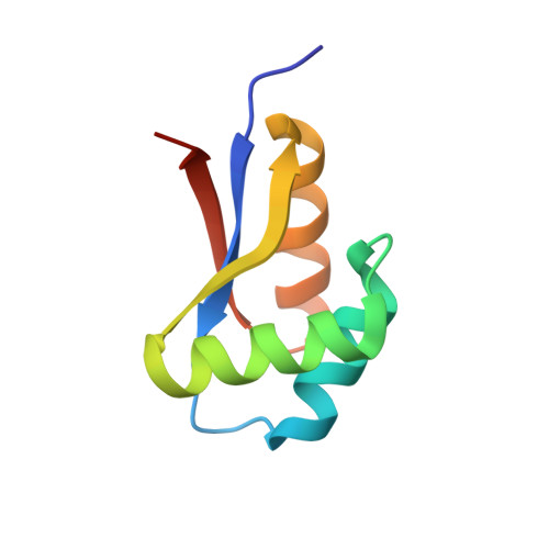
Structural basis for the N-degron specificity of ClpS1 from Arabidopsis thaliana.

Kim, L.Heo, J.Kwon, D.H.Shin, J.S.Jang, S.H.Park, Z.Y.Song, H.K.

(2021) Protein Sci 30: 700-708

  • DOI: https://doi.org/10.1002/pro.4018
  • Primary Citation Related Structures: 
    7D34

  • PubMed Abstract: 

    The N-degron pathway determines the half-life of proteins in both prokaryotes and eukaryotes by precisely recognizing the N-terminal residue (N-degron) of substrates. ClpS proteins from bacteria bind to substrates containing hydrophobic N-degrons (Leu, Phe, Tyr, and Trp) and deliver them to the caseinolytic protease system ClpAP. This mechanism is preserved in organelles such as mitochondria and chloroplasts. Bacterial ClpS adaptors bind preferentially to Leu and Phe N-degrons; however, ClpS1 from Arabidopsis thaliana (AtClpS1) shows a difference in that it binds strongly to Phe and Trp N-degrons and only weakly to Leu. This difference in behavior cannot be explained without structural information due to the high sequence homology between bacterial and plant ClpS proteins. Here, we report the structure of AtClpS1 at 2.0 Å resolution in the presence of a bound N-degron. The key determinants for α-amino group recognition are conserved among all ClpS proteins, but the α3-helix of eukaryotic AtClpS1 is significantly shortened, and consequently, a loop forming a pocket for the N-degron is moved slightly outward to enlarge the pocket. In addition, amino acid replacement from Val to Ala causes a reduction in hydrophobic interactions with Leu N-degron. A combination of the fine-tuned hydrophobic residues in the pocket and the basic gatekeeper at the entrance of the pocket controls the N-degron selectivity of the plant ClpS protein.


  • Organizational Affiliation
    • Department of Life Sciences, Korea University, Seoul, South Korea.

Macromolecule Content 

  • Total Structure Weight: 18.1 kDa 
  • Atom Count: 1,240 
  • Modeled Residue Count: 158 
  • Deposited Residue Count: 164 
  • Unique protein chains: 1

Macromolecules

Find similar proteins by:|  3D Structure
Entity ID: 1
MoleculeChains  Sequence LengthOrganismDetailsImage
ATP-dependent Clp protease adapter protein CLPS1, chloroplastic
A, B
82Arabidopsis thalianaMutation(s): 0 
Gene Names: CPLS1CLPTAt1g68660F24J5.10F24J5.4
UniProt
Find proteins for Q9SX29 (Arabidopsis thaliana)
Explore Q9SX29 
Go to UniProtKB:  Q9SX29
Entity Groups
Sequence Clusters30% Identity50% Identity70% Identity90% Identity95% Identity100% Identity
UniProt GroupQ9SX29
Sequence Annotations
Expand
Reference Sequence

Experimental Data & Validation

Experimental Data

  • Method: X-RAY DIFFRACTION
  • Resolution: 2.01 Å
  • R-Value Free:  0.299 (Depositor), 0.299 (DCC) 
  • R-Value Work:  0.274 (Depositor), 0.274 (DCC) 
  • R-Value Observed: 0.277 (Depositor) 
Space Group: P 21 21 2
Unit Cell:
Length ( Å )Angle ( ˚ )
a = 81.056α = 90
b = 38.005β = 90
c = 61.411γ = 90
Software Package:
Software NamePurpose
PHENIXrefinement
HKL-2000data reduction
HKL-2000data scaling
PHENIXphasing

Structure Validation

View Full Validation Report


Ligand Structure Quality Assessment 


Entry History 

Deposition Data

Revision History  (Full details and data files)

  • Version 1.0: 2021-04-28
    Type: Initial release
  • Version 1.1: 2023-11-29
    Changes: Data collection, Database references, Refinement description